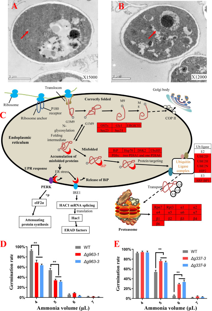
FIG 3.
Analyses of ER stress induced by ammonia fungistasis. (A and B) Ultrastructural analyses of fresh conidia (A) and fungistatic conidia (B) by transmission electron microscopy; the arrows indicate the possible area of ER. (C) Distribution of up-expressed proteins in “proteasome,” “protein processing in endoplasmic reticulum,” and “ubiquitin ligase complex”; up-expressed proteins are shown with a red background. (D) Germination rate testing of gene AOL_s00054g963 knockout mutants. (E) Germination rate testing of gene AOL_s00210g337 knockout mutants. WT, wild-type strain. Δg963-1 and Δg963-3, knockout mutants of gene AOL_s00054g963. Δg337-3 and Δg337-9, knockout mutants of gene AOL_s00210g337. **, P < 0.01.

