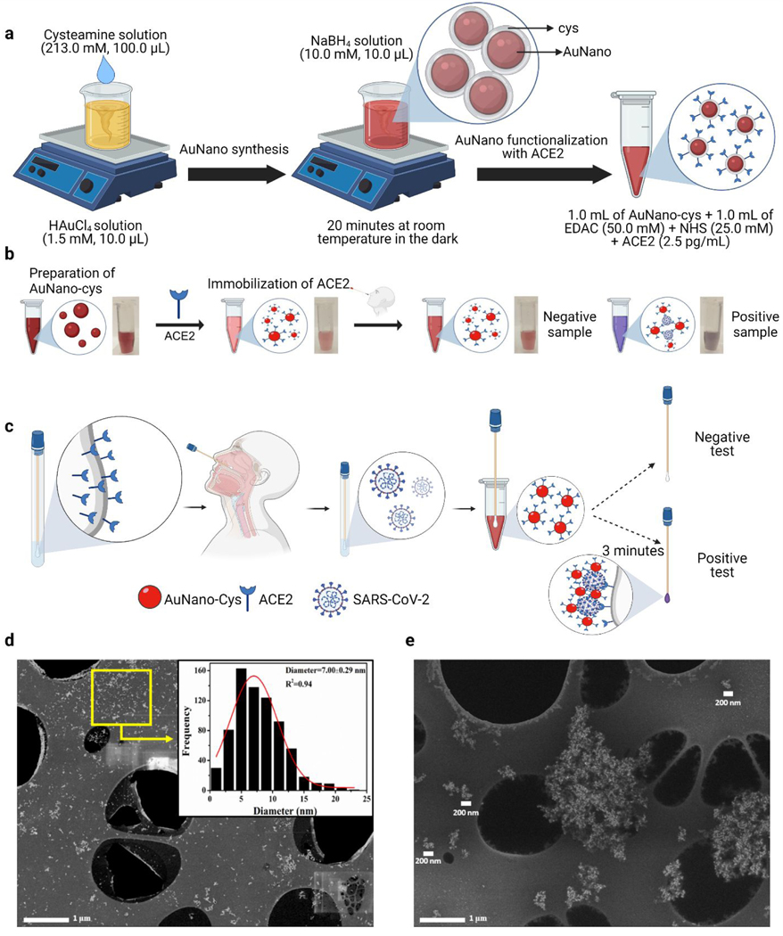
Figure 1. Design, manufacturing, and characterization of COLOR.
(a) Schematic representation of the synthesis steps needed to generate AuNano, including their functionalization with cysteamine (cys) and ACE2. Briefly, cys was added in the presence of chloroauric acid for 20 min at room temperature and protected from light. Sodium borohydride was then added to the solution at room temperature in a light-protected flask. Next, the AuNano-cys were exposed to a mixture of ACE2 and EDAC:NHS for functionalization of the enzyme on the AuNano-cys surface. (b) Steps and photos showing the colorimetric detection of SARS-CoV-2 in an aqueous medium using the synthesized AuNano-cys-ACE2. Proof-of-principle methodology for recognition of the SARS-CoV-2 spike protein through color change (from red to purple, due to the plasmonic effect) of the gold nanoparticles upon aggregation. (c) Schematic representation of the colorimetric steps needed for COVID-19 diagnosis using COLOR. In the presence of SARS-CoV-2, a color shift occurs, and the cotton swabs change color from white to purple after 3 minutes. (d) Morphological characterization of the AuNano-cys solution, which presented a spherical shape and a high dispersion. Inset shows a histogram depicting the size distribution for AuNano-cys with a mean diameter size of 7.00 nm. (e) SEM micrograph of AuNano-cys-ACE2 aggregated in the presence of SARS-CoV-2 SP. Scale bars illustrate that clusters formed between the AuNano-cys-ACE2 and the virus are larger than 200 nm.

