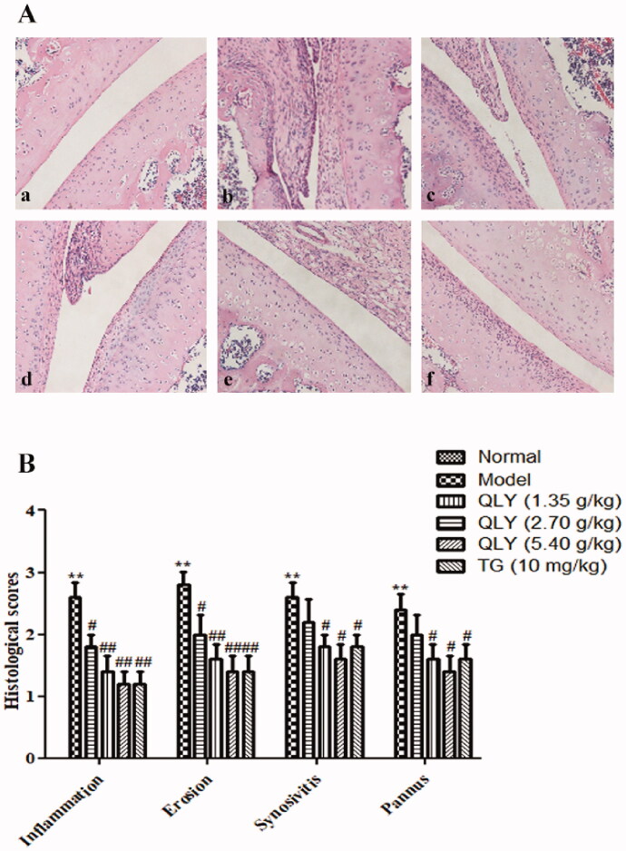
Figure 4.
Effects of QLY granules on histopathology of AA joints. The histopathology examinations in joints were observed by H&E staining. (A) Representative histological changes of haematoxylin and eosin-stained sections of the joints (magnification × 400). a: normal; b: model; c: QLY granules (1.35 g/kg); d: QLY granules (2.70 g/kg); e: QLY granules (5.40 g/kg); f: TG (10 mg/kg). (B) Histopathological evaluation of the synovium from the AA rats. The histological appearance was scored for the presence of synovial proliferation, infiltrated inflammatory cells, pannus formation, and cartilage erosion. Data are expressed as the mean ± SD, with 5 animals in each group. **p < 0.01 vs. normal; #p < 0.05, ##p < 0.01 vs. model.

