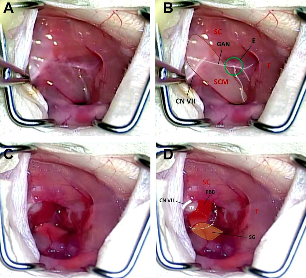
Fig. 2.
(A, B) The surgical anatomy of the neck after the skin incision and retraction of underlying fibromuscular tissue. CN VII = facial nerve; E = Erb’s point; GAN = greater auricular nerve; SC = splenius capitis muscle; SCM = sternocleidomastoid muscle; T = trapezius muscle. (C, D) The GAN, SCM, and anterior scalene muscle have been divided, exposing the proximal extratemporal portion of CN VII. This dissection reveals the PBD. PBD = posterior belly of digastrics muscle; SG = submandibular gland; T = trapezius muscle; TB = tympanic bulla.

