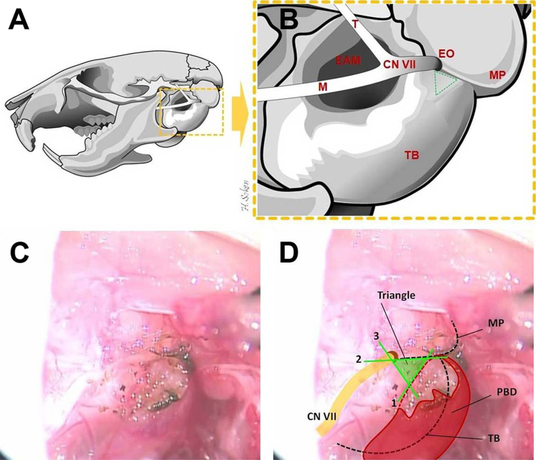
Fig. 3.
Temporal bone anatomy and the key landmarks for placement of the tympanotomy. (A, B) The left mouse temporal bone, lateral view, is illustrated. EAM = external acoustic meatus; EO = external orifice of facial nerve (stylomastoid foramen); M = mandibular branch of the facial nerve; MP = mastoid process; T = temporalis branch of the facial nerve; TB = tympanic bulla. (B, C, D) The lines forming the “Tympanotomy Triangle” are both illustrated and depicted in vivo as follows: (1) anterior border of the PBD insertion and/or its bony ridge (digastric ridge), (2) the fissure between TB and MP (tympanomastoid fissure), (3) the line joining the two by crossing the EO (stylomastoid foramen). PBD = posterior belly of digastrics muscle.

