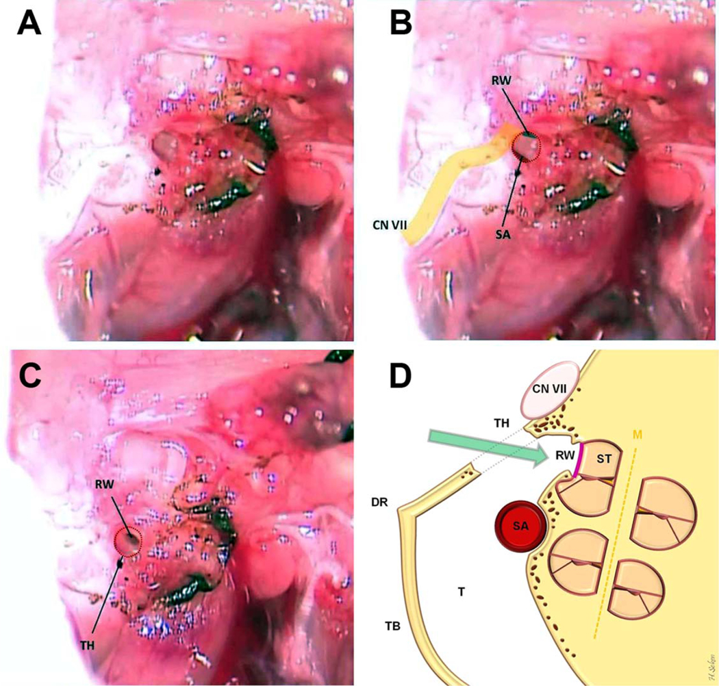
Fig. 4.
Access to the middle ear from our tympanotomy hole is shown (A, B). The inferior margin of RW and superior margin of SA are clearly visible. (C) The final view of the tympanotomy hole when complete and ready for RW implantation. (D) The coronal illustration of the cochlea and surgical preparation. CN VII = facial nerve; DR = digastric ridge; M = modiolus; RW = round window; SA = stapedial artery; ST = scala tympani; T = tympanum; TB = tympanic bulla; TH = tympanotomy hole.

