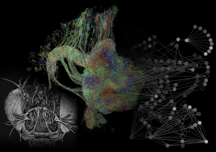
Figure 1. The connectome of the central complex of a fruit fly.
Using electron microscopy, the brain of the fruit fly (Drosophila melanogaster, head depicted on the left) was imaged at high resolution. All neurons of the central complex were reconstructed (some of which are shown in the middle image) and their synaptic connections were extracted. The resulting neuronal networks (symbolically illustrated on the right) were linked to the functions they have in controlling the fly’s behavior.
Image credit Heinze 2021 (CCBY 4.0). Middle image obtained via https://hemibrain.neuronlp.fruitflybrain.org/.

