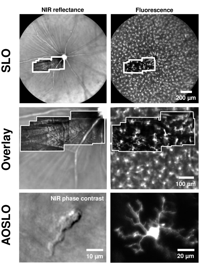
Fig. 1.
Adaptive optics (AO) resolves details of single microglial cell processes in vivo, in NIR label-free and fluorescence channels. Top: Wide-field images of the retina captured with a commercial SLO, with NIR confocal reflectance (left) and EGFP fluorescence (right) in a CX3CR1-GFP mouse. Overlaid inset (white border) shows montage of AOSLO dual-channel images from the same mouse (with simultaneous collection of confocal reflectance and fluorescence). Middle: Zoom-in of top images, to compare SLO images with overlaid AOSLO montages from the same mouse (white border). AOSLO offers much higher axial sectioning, therefore isolating fluorescence from a single stratification of microglia, unlike the SLO. Bottom: High-resolution AOSLO images (single fields) showing microglial cell soma and its processes, in both label-free NIR phase contrast (left) and fluorescence (right). (Bottom panel shows two different locations. Figure 8 shows simultaneous the CX3CR1-GFP fluorescence from same cell imaged in the label free configuration on the left.)

