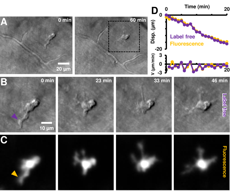
Fig. 9.
Label-free imaging of EGFP+ cell process motility. A) Label-free imaging with near-infrared light (796 nm) using phase-contrast AOSLO. An EGFP+ cell is observed in the top-right quadrant of the image; and confirmed with positive CX3CR1 labelling in C. The cell is tracked in vivo for ∼1 hour. B) Zoom-in of dotted black box region in A, shown at four representative times. Complete time-lapse (10 second resolution) shown in Visualization 7 (201.6MB, avi) . Cell soma and processes can be visualized without use of any exogenous contrast agents. C) Simultaneously imaged fluorescence showing positive CX3CR1 labelling. Cell processes visible are visible in both the infrared label-free and visible channels, showing the potential use of our label-free approach for noninvasive imaging in both mice and humans. Power levels at cornea: label-free 796 nm: 212 µW, fluorescence 488 nm ex.: 53 µW. D) Measured process displacement (disp.) as a function of time, measured independently in the label-free and fluorescence channels. The cell-process tracked is shown with arrowheads in B-C, purple (label-free) and dark-yellow (fluorescence). Instantaneous velocity (V) of the process is similarly shown on the bottom panel.

