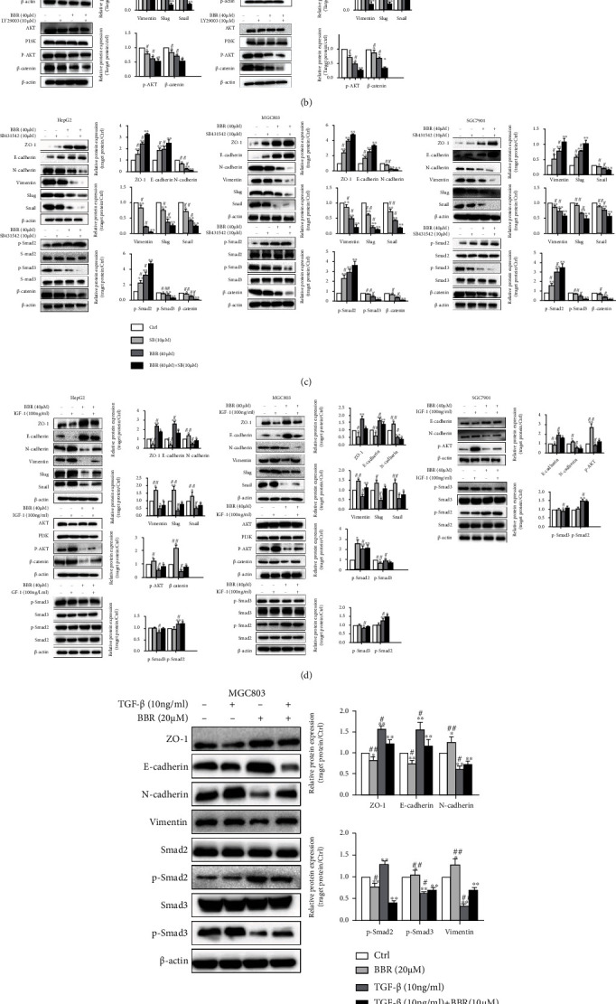
Figure 5.

Effects of BBR on the TGF-β/Smad, PI3K/Akt, and Wnt/β-catenin pathways in HepG2, MGC803, and SGC7901 cells by Western Blot. (a) HepG2, MGC803, and SGC7901 cells were treated with BBR (10, 20, and 40 μM) for 24 h. The pathway proteins of TGF-β/Smad, PI3K/Akt, and Wnt/β-catenin, including Smad2, p-Smad2, Smad3, p-Smad3, Akt, p-Akt, PI3K, and β-catenin, were measured. (b) HepG2 and MGC803 cells were treated alone or cotreated berberine (40 μM) and LY. Measured the proteins of E-cadherin, ZO-1, N-cadherin, vimentin, Snail, Slug, Akt, p-Akt, PI3K, and β-catenin. (c) Treated alone or cotreated BBR (40 μM) and SB (10 μM), the proteins of E-cadherin, ZO-1, N-cadherin, vimentin, Snail, Slug Smad2, p-Smad2, Smad3, and p-Smad3. (d) and (e) Treated alone or cotreated BBR (40 μM) and IGF-1 (100 ng/ml) or TGF-β (10 ng/ml) the proteins of relating to EMT, Smad2, Smad3, p-Smad2, and p-Smad3 and were measured. (f) To compare the effect of BBR (20 μM) and SB (20 μM) in TGF-β pathway, Smad2, Smad3, p-Smad2, and p-Smad3 protein expression level was detected. β-Actin was used as a loading control. Representative images and typical graphs (mean ± SD) are shown, (n = 3. ∗p < 0.05, ∗∗p < 0.01 versus the control group, #p < 0.05, ##p < 0.01 versus the inhibitors group).
