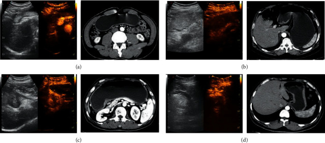
Figure 1.

DCEUS image of typical cases. Note: in (a), (b), (c), and (d), the left images were oral gastrointestinal contrast agent ultrasound images, the middle images were double-contrast ultrasound images, and the right images were enhanced MSCT images. (a) An ultrasound image of a 59-year-old male patient; he was finally diagnosed as a moderately differentiated adenocarcinoma of the antrum (pT1 staging). DCEUS showed focal thickening of the gastric wall, with focal positive development of the middle layer in the inner layer of the gastric wall and clear boundary with the outer layer, and it was judged that the tumor infiltrated into the muscularis mucosa. MSCT showed focal gastric wall thickening, and the lesion did not exceed the low-density zone of submucosa (preoperative diagnosis cT1 staging). (b) An ultrasound image of a 60-year-old female patient. She was finally diagnosed as poorly differentiated adenocarcinoma with ulcerative gastric antrum (pT2 staging). DCEUS showed that the whole gastric wall of the lesion was thickened and positively developed, and the outer edge of the lesion was intact and smooth, and it was judged that the tumor infiltrated into the muscle layer; MSCT showed that the gastric wall was thickened, the lesion broke through the slightly strengthened muscle layer of the submucosa, the outer surface of the stomach around the lesion was clear and smooth, and the fat surface around the stomach was clear (preoperative diagnosis of cT2 staging). (c) An ultrasound image of a 64-year-old female patient. She was finally diagnosed as a poorly differentiated adenocarcinoma with an ulcerative type of lesser curvature of stomach (pT3 staging). DCEUS showed that the whole gastric wall of the lesion was obviously thickened and showed positive development, and the outer layer of the tumor was vague and serrated, breaking through the adventitia, but not invading the adjacent structures, and it was judged that the tumor infiltrated into the subserosal layer; MSCT showed gastric wall thickening, irregular fat infiltration around the stomach, and uneven serosal surface (preoperative diagnosis of cT3 staging). (d) An ultrasound image of a 58-year-old male patient. He was finally diagnosed as poorly differentiated adenocarcinoma of the lesser curvature ulcerative type (pT4 staging). DCEUS showed that the whole layer of gastric wall of the lesion was thickened and showed positive development, the tumor invaded the outer serous, and the tumor broke through serosa; MSCT showed that the tumor broke through the perigastric adipose tissue and serosa, accompanied by the expansion and invasion of adjacent organs or structures (preoperative diagnosis of cT4 staging).
