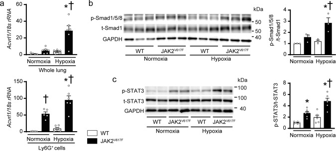
Fig. 6. Acvrl1 mRNA expressions and phosphorylation of Smad1/5/8 and STAT3 in the lungs of JAK2V617F mice in response to chronic hypoxia.
a mRNA expression of Acvrl1 in whole lung extracts (top) or the sorted Ly6G+ cells from the lungs (bottom) of WT mice and JAK2V617F mice exposed to normoxia or hypoxia. The data were normalized to 18 s rRNA levels (n = 5, 6, 5, 6, *P = 0.0004, †P = 0.0004 for whole lung extracts, n = 5, 5, 6, 6, *P = 0.0069, †P = 0.0012 [left], <0.0001 [right] for sorted Ly6G+ cells). Western blot analysis on the SMAD (b) and STAT (c) pathways in the lungs. Lung extracts from WT mice or JAK2V617F mice were immunoblotted with the indicated antibodies. The ratios of phosphorylated Smad1/5/8 (p-Smad1/5/8) to total Smad1 (t-Smad1) and phosphorylated-STAT3 (p-STAT3) to total STAT3 (t-STAT3) are shown in the bar graphs. The average value for normoxia-WT mice was set to 1 (b, n = 3 in each group, *P = 0.0382, †P = 0.0100; c, n = 6 in each group, *P = 0.0125 [left], 0.0019 [right], †P < 0.0001). GAPDH was used as the loading control. All data are presented as mean ± SEM. *P < 0.05 versus the corresponding normoxia-group and †P < 0.05 versus the corresponding WT mice by the one-way ANOVA with Tukey post-hoc analysis. Source data are provided as a Source Data file.

