Figure 1.
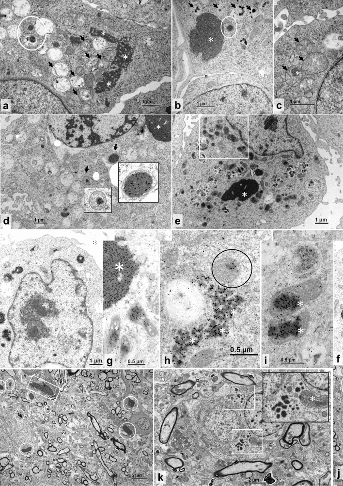
TEM and immuno-EM of reptarenavirus-infected and NP-transfected boid cells, and TEM of a BIBD-positive B. constrictor brain. (a–c) TEM, permanent cell culture derived from B. constrictor kidney (I/1Ki), infected with UGV-1, at three dpi. (a) Large irregular cytoplasmic inclusion body (IB; asterisks), vacuolated mitochondria (arrows) and partly ruptured mitochondrion with electron-dense IBs in the matrix (circle). (b) Large electron-dense cytoplasmic IB (asterisk), one mitochondrion with IB in the matrix (circle), and several vacuolar structures consistent with vacuolated mitochondria with small IBs (arrows). (c) Swollen mitochondria with finely granular disintegrated matrix (arrows) and one IB (asterisk). (d) Immunogold labelling of the reptarenaviral NP, I/1Ki infected with UGV-1, at three dpi. Large NP-positive cytoplasmic IB (asterisk) and several NP-positive IBs within the matrix of mitochondria (arrows). Inserts: higher magnification of the areas indicated by the arrows. (e–i) I/1Ki transfected with UHV1-NP-FLAG, at three dpt. (e) TEM, cell with large electron-dense IB (asterisk) and multiple mitochondria with IB formation (highlighted by a white rectangle). (f–i) Immunogold labelling of reptarenaviral NP. (f) Positive reaction in small electron-dense cytoplasmic IBs (asterisks). (g) Large electron-dense IB with positive reaction (larger asterisk) and individual mitochondria with positive IBs within the matrix (smaller asterisks). (h) Irregular shaped, more electron-lucent, presumably earlier cytoplasmic IB (asterisk) and single mitochondrion with positive reaction within the matrix (circle). (i) Mitochondria with positive IBs within the matrix (asterisks) at higher magnification. (j–k) TEM, neurons. (j) Numerous < 1–3.5 µm sized, round, smooth edged electron-dense cytoplasmic IBs (circles) and one larger, less electron-dense, irregularly shaped IB with coarser margins (square). (k) Neurons with small, 0.1–3 µm sized electron-dense cytoplasmic IBs (white squares). Insert: higher magnification depicting irregularly shaped IB borders and a swollen mitochondrion with disorganized coarse electron-dense matrix (asterisk).

