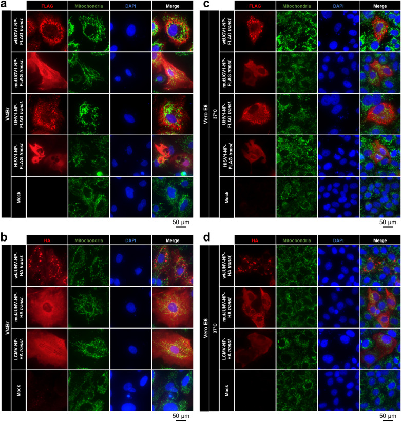
Figure 6.
IF studies on arenaviral NPs in transfected boid and mammalian cells. Double IF images of Boa constrictor V/4Br cells incubated at 30 °C (a,b) or of monkey Vero E6 cells incubated at 37 °C (c,d), transfected with a construct expressing either wt or mutUGV1-NP-FLAG, UHV1-NP-FLAG, or HISV1-NP-FLAG (a,c), and wt or mutJUNV-NP-HA, or LCMV-NP-HA (b,d) at three dpt. Non-transfected (Mock) cells served as controls. (a,c) The panels from left: FLAG tag in red (secondary antibody: AlexaFluor 594 goat anti-rabbit), mitochondrial marker in green (secondary antibody: AlexaFluor 488 goat anti-mouse), nuclei in blue (DAPI), and a merged image. (b,d) The panels from left: HA tag in red (secondary antibody: AlexaFluor 594 goat anti-rabbit), mitochondrial marker in green (secondary antibody: AlexaFluor 488 goat anti-mouse), nuclei in blue (DAPI), and a merged image.

