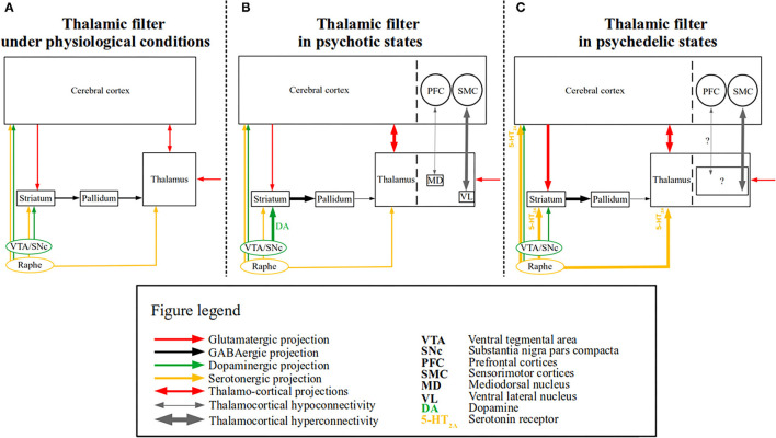
Figure 1.
The “thalamic filter” model in psychotic and psychedelic states. A simplified schematic of the “thalamic filter” model is depicted along with the associated thalamocortical dysconnectivity in psychotic and psychedelic states. (A) Depicts thalamic filtering under physiological conditions, in which thalamic activity is modulated by several sources (e.g., cortico-striatal), which are in turn modulated by distinct neurotransmitter systems. Glutamatergic (red arrows), GABAergic (black arrows), dopaminergic (green arrows), and serotonergic projections (yellow arrows) are shown. Cortico-striatal projections are depicted by a unidirectional red arrow from the cerebral cortex to the striatum, whereas the reciprocal projections between the thalamus and the cerebral cortex are depicted by a bidirectional red arrow. Finally, incoming (glutamatergic) sensory input to the thalamus is depicted by a red unidirectional arrow. (B) Depicts a proposed model of altered thalamic filtering in psychotic states, induced via aberrant dopamine function, based on the model provided by Carlsson et al. (19). The left side shows the impaired thalamic filter function, which is induced by excess dopamine (shown as DA) and increased dopaminergic signaling (depicted by the thick green arrow), which leads to an increased inhibition of the pallidum via the striatum (depicted by the thick black arrow) and reduced thalamic inhibition via the pallidum (depicted by the thin black arrow). The enhanced thalamic disinhibition (i.e., “opening” of the thalamic filter) results in sensory flooding (depicted by the thick bidirectional red arrow between the thalamus and the cerebral cortex), presumably bringing about (psychotic) symptoms [for similar models see (20, 21)]. On the right side, separated by the dashed line both in the cerebral cortex and thalamus, rsfMRI findings of thalamocortical dysconnectivity in psychotic disorders are summarized [e.g., (22, 23)]. Thalamocortical hyperconnectivity (depicted by the thick bidirectional gray arrow) between the ventrolateral thalamus (VL) and sensorimotor cortices (SMC) and hypoconnectivity (depicted by the thin bidirectional gray arrow) between mediodorsal thalamus (MD) and prefrontal cortices (PFC), are shown. (C) Depicts a proposed model of altered thalamic filtering in psychedelic states, slightly modified from (9, 18). The left side shows the impaired thalamic filter function induced by psychedelic stimulation of 5-HT2A receptors, which leads to increased extracellular glutamate levels in the prefrontal cortex and inhibitory activity of interneurons in the basal ganglia, thereby increasing the excitatory effect of pyramidal neurons both on the basal ganglia (depicted by the thick unidirectional red arrow between the cerebral cortex and the striatum) and the thalamus (depicted by the thick bidirectional red arrow between the cerebral cortex and the thalamus) (24, 25). Recent evidence also indicates that psychedelics (i.e., LSD) modulate activity in the reticular thalamus leading to the disinhibition of other thalamic nuclei (i.e., mediodorsal nucleus—not shown) (26). In parallel, the increased striatal activity leads to a similar chain of events as seen in psychotic states (i.e., also resulting in enhanced thalamic disinhibition) (18). On the right side, separated by the dashed line both in the cerebral cortex and thalamus, rsfMRI findings of thalamocortical dysconnectivity in psychedelic states are summarized [e.g., (27, 28)]. The driving thalamic regions involved in these phenomena have not yet been identified (depicted by the black square containing “?”), nor is it clear whether thalamocortical hypoconnectivity (thin gray bidirectional arrow with “?”) is present.

