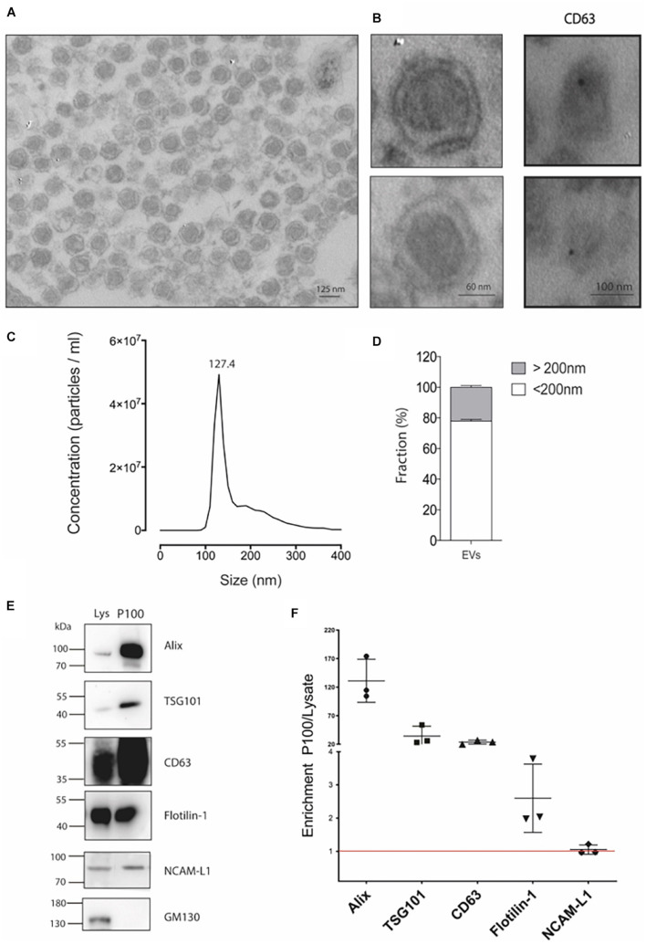
FIGURE 1.
Detailed characterization of P100 vesicles secreted from hippocampal HT-22 cells. (A) Representative transmission electron microscopy of P100 shows abundant vesicles with exosome-like appearance with an average diameter of 124.1 ± 6.8 nm of (n = 31; three independent cell culture preparations). (B) Close capture of single exosomes (left) and immunogold labeling of a selective exosomal biomarker, CD63 (right panel, black dot). (C) Nanoparticle tracking analyses show a mean peak at 127.4 ± 1.3 nm (n = 5; five independent cell culture preparations). (D) Abundance analysis shows that 77.88 ± 2.41% of the vesicles have a diameter of <200 nm and a 22.12 ± 2.41% >200 nm (n = 4; four-five independent cell culture preparations). (E,F) Western blot and Analysis enrichment (fold) of the protein content of the exosomal markers Alix, 131.08 ± 37.7; TSG101, 34.36 ± 17.00; CD63, 23.82 ± 3.50; Flotillin-1, 2.60 ± 1.03; NCAM-L1, 1.05 ± 0.14. The control GM130, a Golgi-associated protein, was absent. Enrichment is relative to their levels in the lysate. The values were obtained from three independent cell culture preparations.

