Table 5.
Chemical properties and biological activity of photosensitizers used in vivo/ex vivo pre-clinical studies for treatment of infections by Gram-negative bacteria
| # | Structure | Model | Results |
|---|---|---|---|
| 1 |
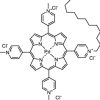
FS111-Pd (PS36) Charge: + 4 MW = 938 Da |
In vivo: adult female BALB/c mice with wound infection by E. coli [64] |
Outcome: 4 log CFU reduction initially. Complete inactivation 4 days after treatment [PS]: 50 µM (50 + 20 + 20 µl) Light dose: 80 J/cm2 (light λ = 415 nm) |
| 2 |
Photogem (PS31) Charge: − 2 to − 12 MW = n.d. |
In vivo: Mongolian Gerbils with otitis caused by H. influenza ATCC 19418 [167] |
Outcome: complete reduction of H. influenza in 50% of infections [PS]: 1 mg/ml (20 μl) Light dose: n.d. total energy: 90 J (laser λ = 632 nm) |
| 3 |
5-ALA: protoporphyrin IX precursor (PS9a) Charge: 0 MW = 131 Da |
In vivo: Kunming mice infected with P. aeruginosa ATCC 27853 [177] |
Outcome: 1 log CFU reduction after treatment [PS]: 1.4 M ALA Light dose: 54 J/cm2 (light λ = 630) |
| 4 |
Verteporfin (PS37) Charge: − 1 MW = 718 Da |
In vivo: male BALB/c mice with subcutaneous Mycobacterium bovis induced granuloma sites [178] |
Outcome: 0.7 log CFU reduction, 72 h after treatment [PS]: 0.5 mg/kg (0.7 µmol/kg) Light dose: 60 J/cm2 (laser λ = 690 nm) |
| 5 |
EtNBSe (PS38) Charge: + 1 MW = 445 Da |
In vivo: male BALB/c mice with subcutaneous M. bovis induced granuloma sites [179] |
Outcome: 2 log CFU reduction [PS]: 5.25 mg/kg (11.8 µmol/kg) Light dose: 60 J/cm2 cm2 (laser λ = 635 nm) |
| 6 |
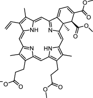
Chlorin e6-polyethylenimine conjugate (PS30) Charge: positive MW = 10,000–25,000 Da |
In vivo: female BALB/c mice wound infected with A. baumannii ATCC BAA 747 [180] |
Outcome: 3 log CFU reduction 30 min after treatment. 1.7 log 1–2 days after [PS]: 800–900 µM (50 µl) Light dose: 240 J/cm2 (light λ = 660 ± 15 nm) |
| 7 |
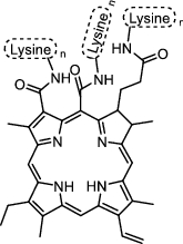
Poly-l-lysine-cp6 (PS39) Charge: positive MW = n.d. |
In vivo: female Swiss albino mice with wound infected with P. aeruginosa MTCC 3541 [181] |
Outcome: 2 log CFU reduction 24 h after treatment [PS]: 200 µM (25 µl) Light dose: 120 J/cm2 (light λ = 660 ± 25 nm) |
| 8 |
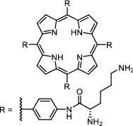
Tetra-lysine porphyrin (PS40) Charge: 0 to + 8 (pH dependent) MW = 1188 Da |
In vivo: Sprague–Dawley rats with wound infected by mixed bacteria (E. coli, S. aureus, P. aeruginosa) [182] |
Outcome: 5 log CFU reduction, 7 days after treatment [PS]: 40 µM Light dose: 100 J/cm2 (laser λ = 650 nm) |
| In vivo: adult female BALB/c mice wound infected with multi-resistant A. baumannii (clinical isolate) [183] |
Outcome: 4 log CFU reduction, 4 days after treatment [PS]: 40 µM Light dose: 50 J/cm2 (Laser λ = 650 nm) |
||
| 9 |
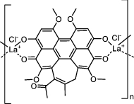
HB: La + 3 (PS41) Charge: positive MW = n.d. |
In vivo: adult female BALB⁄c Mice with burns infected by P. aeruginosa (clinical isolate) [184] |
Outcome: 2 log CFU reduction in bacteria recovered from blood [PS]: 10 µM (100 µl) Light dose: 24 J/cm2 (LED λ = 460 ± 25 nm) |
| 10 |
SACUR-3 (PS35) Charge: + 4 MW = 517 Da |
Ex vivo: porcine skin infected with E. coli ATCC 25922 [176] |
Outcome: 3 log CFU reduction [PS]: 50 µM Light dose: 34 J/cm2 (LED λ = 435 ± 10 nm) |
| 11 |
MB-PMX (PS25) Charge: + 1 MW = 1723 Da |
Ex vivo: porcine skin infected with E. coli ATCC 25922 [154] |
Outcome: 7 log CFU reduction [PS]: 50 µM Light dose: 288 J/cm2 (LED λ = 625 nm) |
| 12 |
Methylene blue (PS18) Charge: + 1 MW = 284 Da |
Ex vivo: cultured human epithelial surfaces infected with MRSA [174] |
Outcome: 5 log CFU reduction [PS]: 300 µM with 0.25% chlorhexidine glucose Light dose: 96 J/cm2 (LED λ = 670 nm) |
| 13 | In vivo: female BALB/c mice infected with cecal slurry [185] |
Outcome: improved wound healing [PS]: 100 µM Light dose: 24 J/cm2 (LED λ = 632 nm) |

