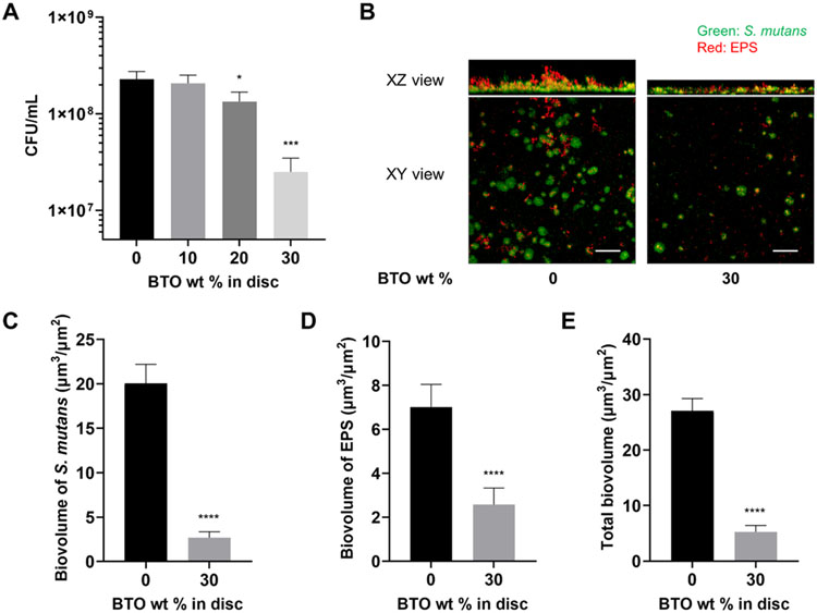
Figure 2.
Antibiofilm activity of BTO-nanocomposite discs. (A) Dose-dependent reduction in CFU/mL with increasing wt% of BTO in discs. Data represent means. Error bars are standard deviations. Statistics: One-way ANOVA: p < 0.001. Post-hoc (Dunnett’s method): *** represents p < 0.001 in comparison to control discs (0 wt%) (n=3). (B) Representative top (XY) and orthogonal (XZ) views of confocal images of S. mutans biofilms at 18 h for control discs (0 wt%) and 30 wt% BTO-nanocomposite discs. Bacterial microcolonies were labeled with SYTO 9 (green) and EPS α-glucan were labeled with Alexa Fluor 647 (red). Scale bar: 50 μm. Quantified biovolume of (C) S. mutans, (D) EPS, and (E) total (sum of S. mutans and EPS) in the biofilm as determined by COMSTAT. Data represent means. Error bars are standard deviations. Statistics: t-test with **** representing p < 0.0001 (n≥3).

