
Fig. 1.
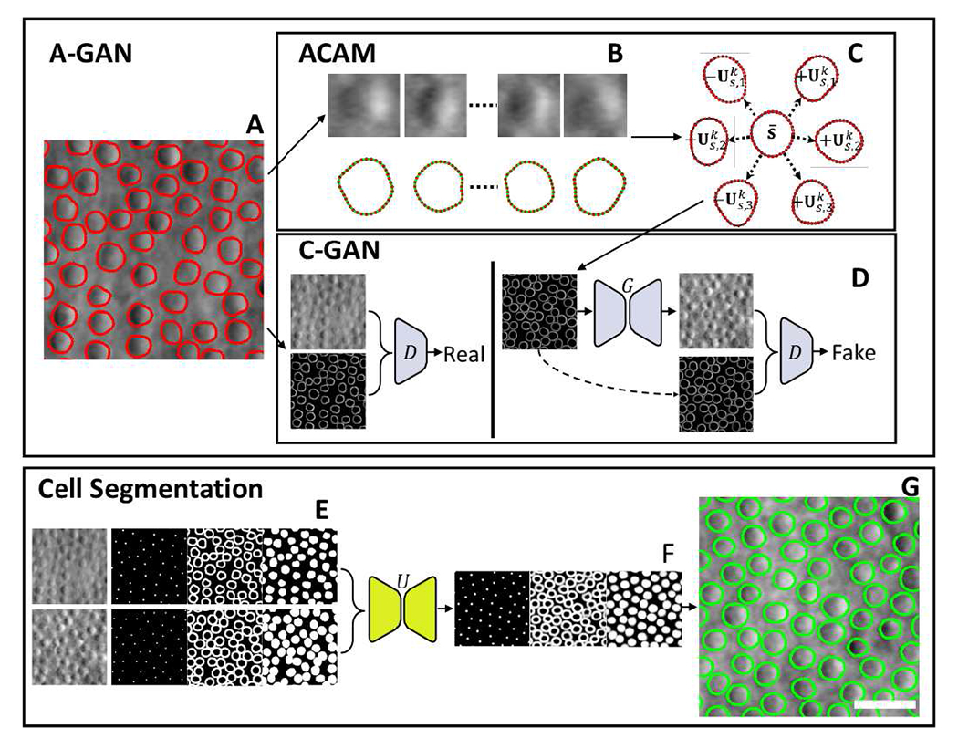
Overview of active cell appearance induced generative adversarial networks (A-GANs) for cell segmentation on (A) adaptive optics retinal images. A-GANs consist of three main steps, including (B) individual cell sample extraction from manual annotations (red contours), (C) active cell appearance model (ACAM), and (D) conditional generative adversarial networks (C-GANs) to create adaptive optics images. (E) The cell segmentation model takes both real and generated adaptive optics images as well as centroid, contour, and region masks as the training data for a U-Net model. The trained U-Net model predicts (F) centroid, contour, and region masks on test images. All of these predicted masks are imported into a level-set segmentation framework to obtain (G) the final cell segmentation (green contours). Scale bar, 20 μm.