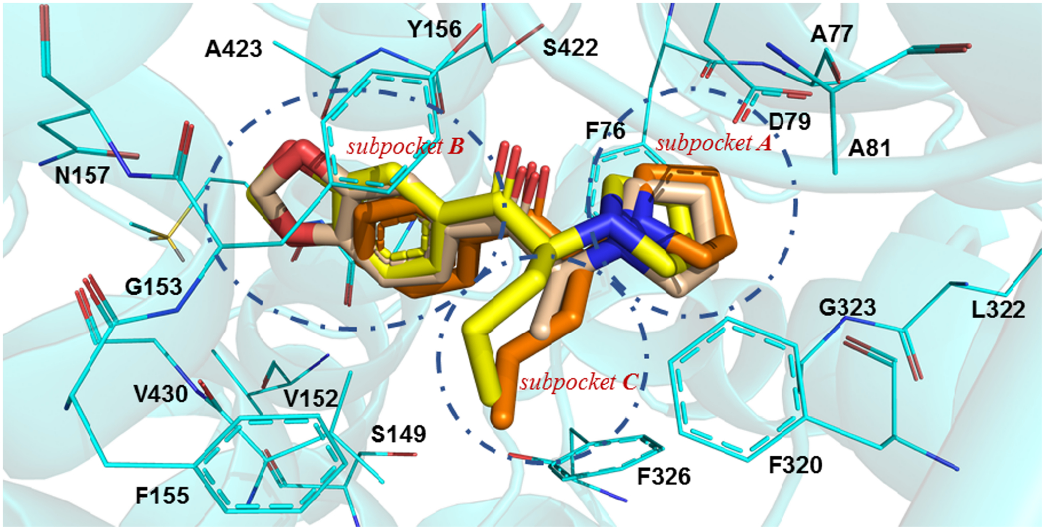
Figure 3.

In silico interaction of MDPV and its analogs at the S1 binding site of hDAT. Possible binding mode of MDPV (capped sticks rendering; yellow carbon atoms), MDPPP (wheat carbon atoms), and α-PVP (orange carbon atoms) at the hDAT homology model (amino acid residues depicted as wire; cyan carbon atoms).
