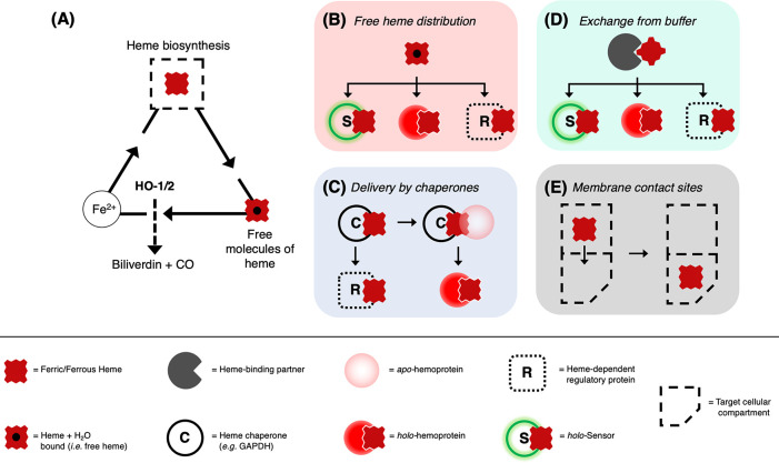
Figure 3.
Representation of possible mechanisms for distribution and delivery of heme across the cell. (A) The life cycle of heme in cells starts with its biosynthesis in the mitochondria (full dashed square) and ends with its degradation by heme oxygenase (HO-1/2). Heme oxygenase generates Fe2+ which can be recycled for the synthesis of new heme molecules. A balance between synthesis and degradation contributes to the controlling heme concentrations in cells. (B–E) The supply of heme to the locations where it is in demand is suggested as occurring via four possible mechanisms (colored panels). In (B), direct distribution of free heme into a heme protein (red circle), a heme-dependent regulatory function (R, white box), or a genetically encoded/synthetic heme sensor for quantification studies (S, green circle). Free heme (either ferrous or ferric) is envisaged as being present in minuscule concentrations but will still represent a mechanism for heme to be made available in cells.77 In (C), chaperone-mediated heme delivery to an apo-heme protein (pale red circle), for example by GAPDH.113−115 In (D), heme bound to heme-binding partners (dark gray pacman) constitutes a body of exchangeable heme readily available for downstream applications, in the same way as in (B). Possible candidates for heme-binding proteins are IDO, HBP22/23, SOUL, and albumin.111,163,196 In (E), distribution of heme via membrane contact sites between mitochondria—where heme is synthesized—to target cellular compartments which bypasses the need for transporters to mediate the delivery of heme.171

