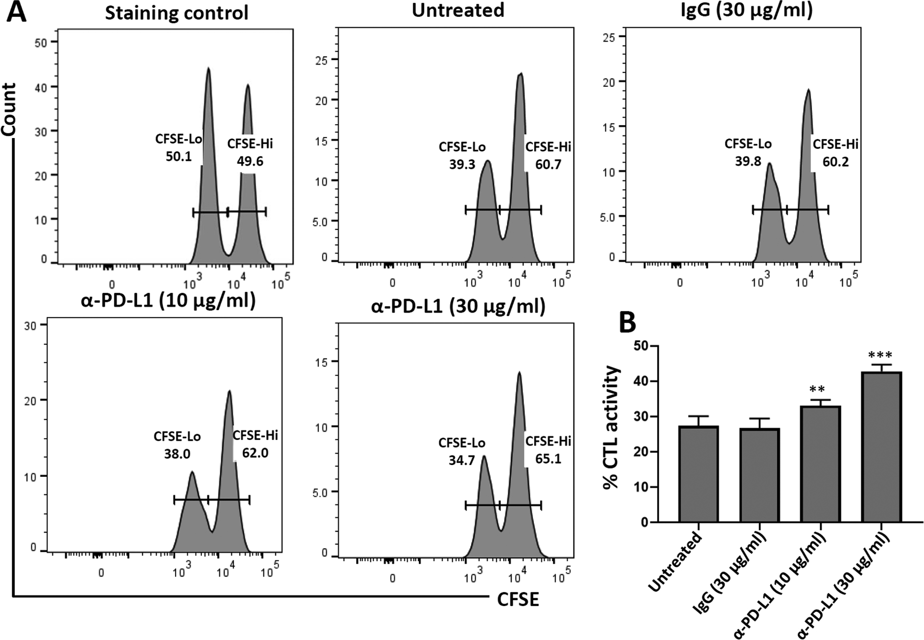
FIGURE 3.

In vitro microglial cell cytotoxicity assays using neutralizing Ab against PD-L1. CD8+ T-cells were isolated from spleens of rAd5-OVA-primed OT-1 mice using a negative selection kit. CD8+ T-cells were then transferred to microglial cells (10:1 effector: target ratio) from C57BL/6 mice loaded with ovalbumin-specific peptide, labeled with CFSE, and either left untreated or treated with IgG control (30 μg/ml) or α-PD-L1 neutralizing Ab (10 μg/ml and 30 μg/ml). in vitro cytotoxicity was determined after 18 hr. (a) Representative histograms show CFSE-labeled microglia harvested from the plate after 18 hr of co-culture with CD8+ T-cells. (b) Graphical representation of % CTL activity [1 − (control group hi/lo ratio: experimental group hi/lo ratio)] × 100. **p < .01; ***p ≤ .001 compared to IgG control. Data shown are representative of three independent experiments
