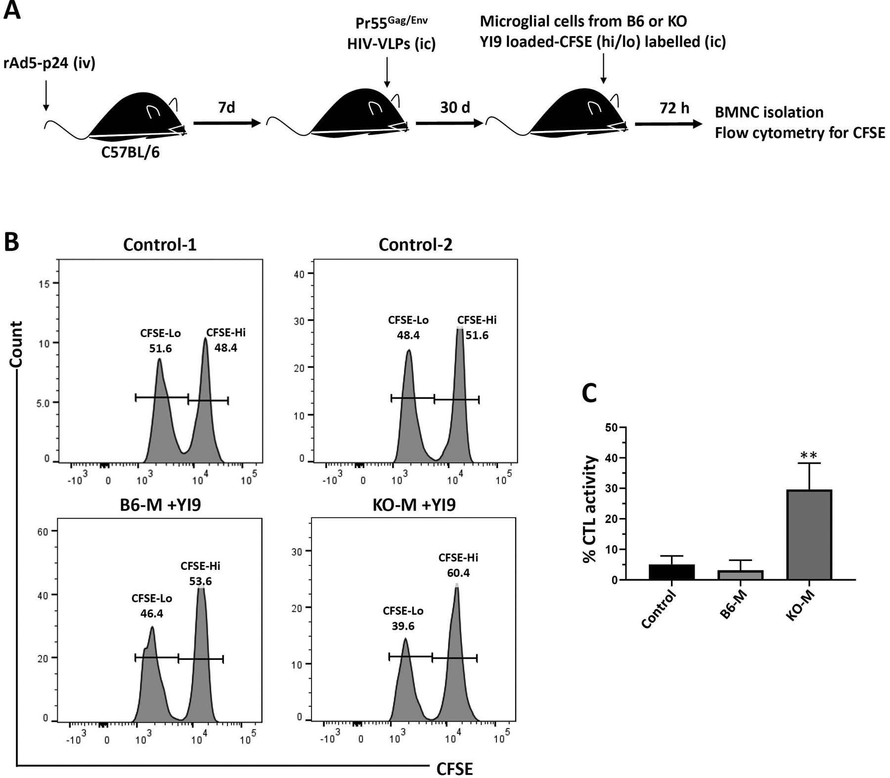
FIGURE 4.

In vivo CTL killing of YI9 peptide-loaded C57BL/6 and PD-L1 KO microglial cells within the brain. (a) C57BL/6 mice were primed via tail-vein injection with rAd5-p24 and boosted in the CNS with HIV-VLPs for 30 days. in vivo brain cytotoxicity was determined 72 hr after intracranial (i.c.) injection of syngenic CFSE-labeled primary microglial cells (0.5 × 10⁶ cells) from either C57BL/6 (B6-M) or PD-L1 KO (KO-M) animals pulsed with or without 1 μg/ml of Gag-specific YI9 peptide. (b) Representative histograms showing CFSE-labeled microglia recovered from the brains of rAd5-p24 primed-CNS boosted hosts. Control-1 is the CFSE staining control representative of either B6-M or KO-M. Control-2 is the representative histogram of either B6-M or KO-M without YI9. (c) Graphical representation of percent specific killing [1 − (control group hi/lo ratio: experimental group hi/lo ratio)] × 100. **p ≤ .01 compared to B6-M as well as the control (B6-M without YI9). Data shown are representative of three experiments using n = 3 animals/group
