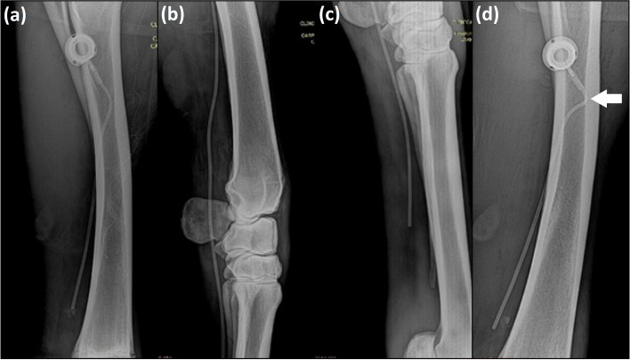
Fig. 1.
Radiographic images of the thoracic limbs of adult horses illustrating in (a): positioning of the 15-cm long totally implantable catheter, model Life-Port Titanium Infant 7.5 Fr in the cephalic vein during the peri-operative period of implantation. In (b): positioning of the 46-cm long totally implantable catheter model Life-Port Titanium Infant 7.5 Fr in the cephalic vein passing through the carpal joint and ending in the middle third of the third metacarpal bone (c). In (d): 24 h after the catheter implantation, standing position; the image shows a steeper curvature of the catheter at the site of coupling to the reservoir (arrow) and the more distal positioning of the reservoir relative to the ulna when compared to the same horse displayed in the image (a)

