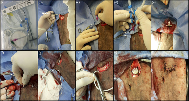
Fig. 7.
Images of the surgical implantation technique of the totally implantable catheter (a) in the cephalic vein of the right thoracic limb of an adult horse: (b) U-shaped skin incision, 10 cm, 3 cm caudal and 3 cm proximal to the determined point for puncture of the cephalic vein; (c) introduction of the puncture needle in the cephalic vein; (d) passage of the “J” shaped guide wire inside the puncture needle and the cephalic vein, followed by the removal of the puncture needle; (e) external portion of the guide wire positioned inside the introducer and dilator and their positioning inside the cephalic vein; (f) after positioning of the introducer and dilator inside the cephalic vein, removing of the guide wire and introducer, followed by the introduction of the totally implantable catheter and removal by “peel off” of the dilator; (g) catheter connected to the tunneling tool and passing through the subcutaneous tunnel created between the site of cephalic vein puncture and elliptical skin incision; (h) after radiographic evaluation, catheter excess being cut to be connect to the reservoir; (i) safe connection of the catheter to the reservoir, correct positioning and placement of the reservoir, suture of the reservoir to the muscle fascia inside the subcutaneous pocket in three points for suture anchorage, proper of the reservoir, with 2–0 nylon; note that the adaptation of the reservoir’s extremity to the catheter should be directed straight to the tunnel through which the catheter was passed, so as not to form curves and/or kinking in the connection between them; (j) suture of the skin over the reservoir and over the puncture point of the cephalic vein, with 0 nylon, simple interrupted suture pattern

