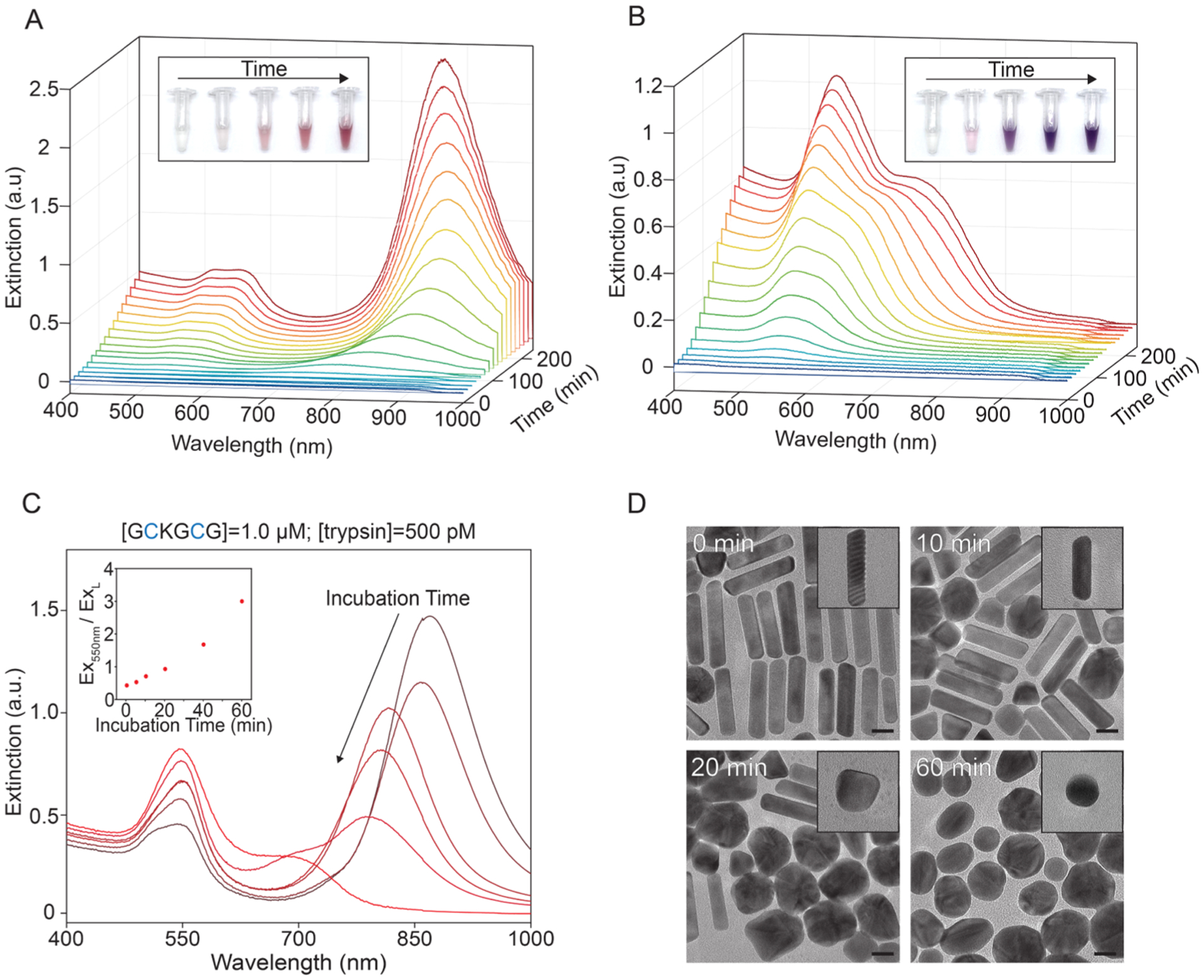
Figure 4.

Temporal study of proteolysis-modulated gold nanoparticle growth. (A,B) UV–vis spectra of gold nanoparticle time evolution with 15 min step size. Inset images were taken 0, 1, 2, 3, and 4 h after seed injection. (A) 1.0 μM GCKGCG and 0 pM trypsin. (B) 1.0 μM GCKGCG and 500 pM trypsin. (C, D) Incubation time-dependent detection of trypsin (Scheme S2). (C) UV–vis spectra of gold nanoparticles prepared with increasing incubation time prior to seed injection: 0, 5, 10, 20, 40, and 60 min. GCKGCG and trypsin concentration held constant. Inset: extinction ratio of Ex550nm to ExL vs incubation time. (D) TEM images of nanoparticles prepared with 1.0 μM GCKGCG and 500 pM trypsin with incubation times of 0, 10, 20, and 60 min; scale bars: 20 nm.
