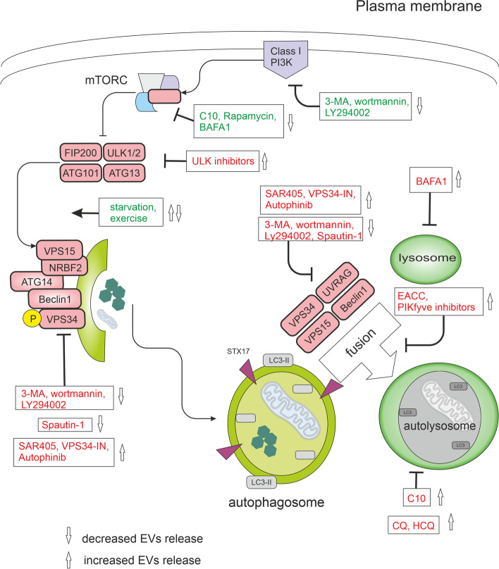
Fig. 7.
Modulators of autophagy and their effect on EVs release. In nutrient-rich conditions, mTORC1 constitutively blocks the ULK complex and autophagy. mTORC1 signals can be inhibited directly by C10, rapamycin, exercise, or starvation and indirectly by bafilomycin A1 (BAFA1) through lysosomal inhibition. Physical exercise was shown to induce the release of small extracellular vesicles (EVs) into the circulation [195]. The ULK complex activates the VPS34 complex. VPS34 is a class III phosphatidylinositol 3-phosphate-kinase (PI3KC3). A group of PI3K inhibitors, including 3-methyladenine (3-MA), wortmannin, and synthetic inhibitor LY294002, inhibits both class I as well as class III PI3Ks. VPS34 inhibitors include Spautin-1, autophinib, SAR405, and VPS34-IN1. Spautin-1 initiates the degradation of Beclin1 due to the inhibition of two of its deubiquitinases. SAR405 and VPS34-IN1 are highly potent inhibitors of VPS34 selective for the VPS34 and not affecting the closely related class I and class II PI3Ks. Autophinib is an ATP-competitive inhibitor of VPS34 decreasing the accumulation of the lipidated protein LC3 on the autophagosomal membrane. The late stages of the autophagic machinery include fusion and degradation. During fusion, the mature autophagosome fuses with lysosomes creating an autolysosome. PIKfyve (phosphoinositide kinase, FYVE-type zinc finger containing) inhibitors and EACC block autophagosome-lysosome fusion. BAFA1 inhibits the acidification of the autolysosome by blocking the V-ATPase while chloroquine (CQ) and 3-hydroxychloroquine (HCQ) impair the maturation of autolysosomes. All drugs are depicted within the rectangles. Effects of modulators activating autophagy are green, inhibitory effects are red

