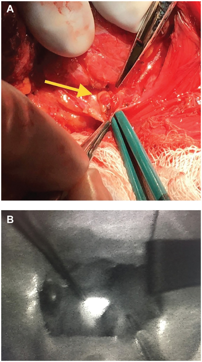
Figure 2.

(A) Normal left inferior parathyroid gland (yellow arrow) from patient 2. (B) Autofluorescence from normal left superior parathyroid gland, seen using the FLUOBEAM LX camera, from patient 5. Both glands corresponded with location on preoperative ultrasound.
