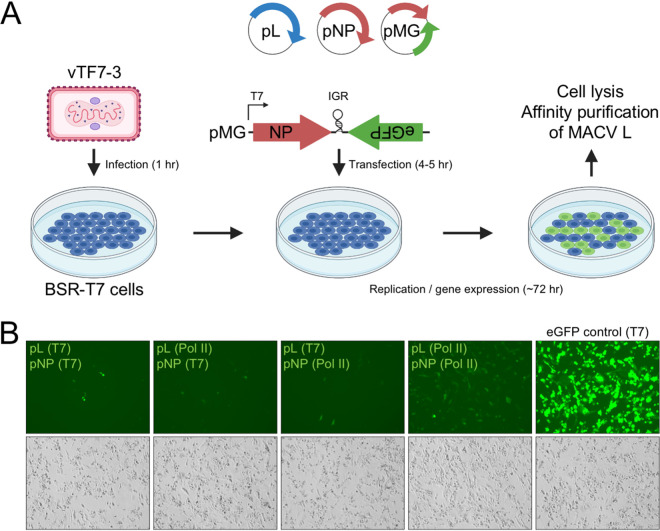
FIG 2.
Expression of affinity-tagged L in the MACV replicon system. (A) Schematic of the MACV minireplicon system. (B) Analysis of minireplicon efficiency for L and NP expressed via different promoters (T7 or Pol II). Fluorescence microscopy images of the eGFP-expressing cells are shown in the upper panel. Bright-field images of infected and transfected cells in the same field of view are shown below. Cells transfected with a plasmid encoding T7 RNAP-driven eGFP are shown in the right-hand column.

