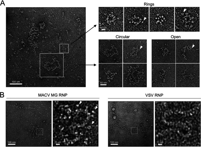
FIG 7.
MACV intracellular RNPs adopt diverse flexible structures. (A) The predominant classifications of MACV RNPs isolated from minireplicon-expressing cells. White outlined boxes highlight the different classes of flexible RNPs observed within a representative negative-stain EM micrograph. Examples of the different classes (rings, circular, and open RNPs) are shown to the right of the micrograph. White arrowheads indicate positions of globular particles on the periphery of the RNPs. (B) Comparison of the MACV RNP filaments with the virion RNP of vesicular stomatitis virus (VSV). White outlined boxes indicate regions of flexible RNP curvature, which are magnified and shown to the right of each micrograph. White arrowheads in the magnified boxes indicate positions of globular particles on the periphery of the RNPs.

