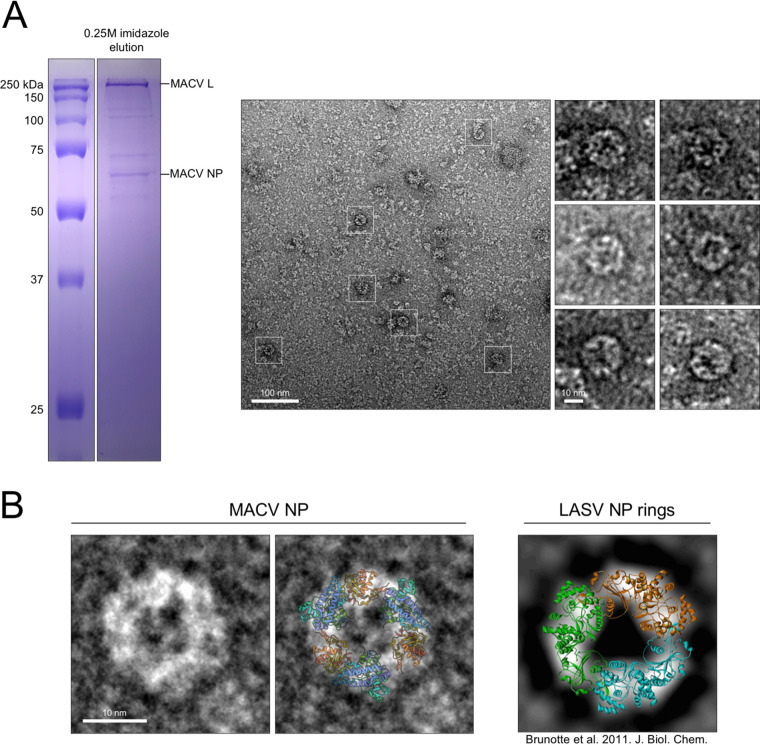
FIG 8.
Intracellular NP trimers associate with L. (A) Polyacrylamide-SDS gel from Fig. 3B shown to highlight the metal ion affinity-purified sample visualized in the negative-stain EM micrograph to the right. White boxes highlight representative ring-like structures, with additional magnified structures shown to the right of the micrograph. (B) Comparison of MACV NP rings to the previously characterized RNA-free rings of LASV (right). A cartoon representation of the LASV NP crystal structure (PDB 3MX5) is overlaid on the MACV ring in the right-hand panel. The LASV NP ring image on the far right is modified from Brunotte et al. (CC-BY 4.0) (51).

