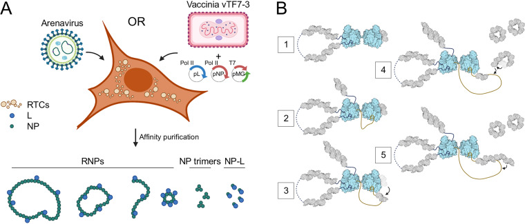
FIG 9.
Model of arenavirus subcellular structures formed by L and NP during infection. (A) Illustration highlighting the diverse structures isolated from MACV minireplicon-expressing BSR-T7 cells. (B) Proposed model for the role of a monomeric L-NP complex in RNP replication. (Step 1) A dimeric MACV L structure is formed with one L subunit (left) serving as the active replicase and the other (right) bound to a single NP monomer waiting to receive the nascent RNA from replicase L. (Step 2) As L begins elongation, the nascent 5′ RNA is bound within the 5′ ligand binding pocket of the adjacent inactive L in a “hand-off” coordinated mechanism, stimulating this polymerase for subsequent RNA synthesis on the corresponding 3′ promoter soon to be synthesized (75). (Step 3) NP-bound L facilitates loading of the NP subunit onto the newly synthesized RNA. (Step 4) Soluble NP trimers are triggered to reverse the gating mechanism of RNA binding, allowing for opening of the ring and engagement of the RNA. (Step 5) NP trimers are loaded onto the growing RNP, either three at a time (depicted here) or one by one.

