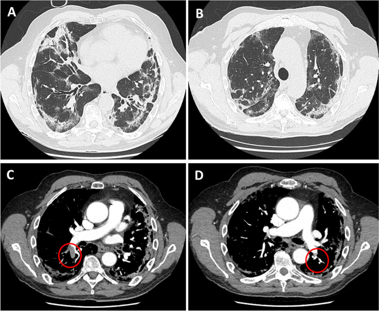
Fig. 1.
Panel A and B Chest CT scan showing diffuse areas of consolidations and interlobular and intralobular septal thickenings involving both lung parenchymas. Panel C and D CT pulmonary angiography in axial view showing filling defects involving the inferior branch of the right pulmonary artery (open circles) and the posterior basal segmental branch of the left pulmonary artery (open circle)

