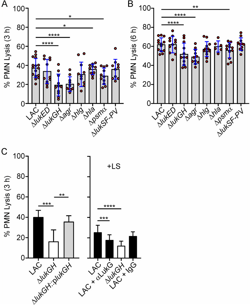
FIG 2.
Lysis of human PMNs following phagocytic interaction with LAC wild-type and isogenic mutant strains. (A, B) PMN lysis 3 h (A) or 6 h (B) after incubation with the indicated strains. Each red circle indicates the results for a separate experiment (n = 10 to 16 [A] and n = 6 to 13 [B]). (C) PMN lysis was determined following phagocytosis of wild-type, ΔlukGH, or complemented ΔlukGH strain (left) or with or without anti-LukG antibody (αLukG) or control IgG as indicated (right) (n ≥ 5). Lysostaphin (LS) was added 30 min after the start of the assay to kill extracellular S. aureus (right) (n ≥ 6). Assays were performed using a 10:1 bacterial cell/PMN ratio, and results are presented as the mean values ± SD. (A, B) Data were analyzed using a mixed-effects analysis and Dunnett’s posttest. (C) Data were analyzed using a mixed-effects analysis and Tukey’s (left) or Dunnett’s (right) posttest. *, P < 0.05; **, P < 0.01; ***, P < 0.001; ****, P < 0.0001.

