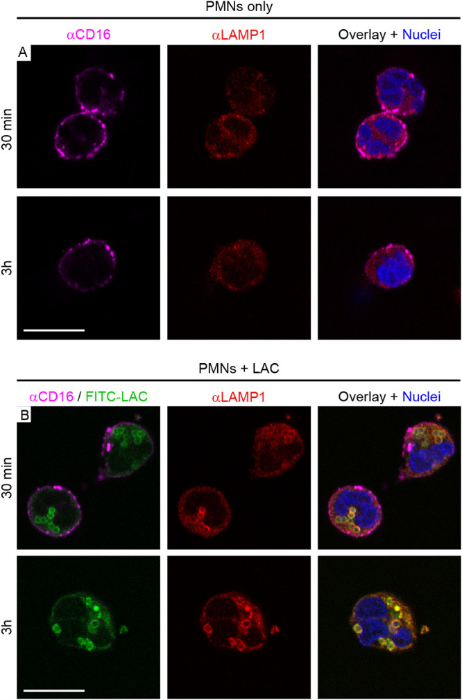
FIG 4.

Subcellular distribution of CD16 and LAMP1 in human PMNs following phagocytosis of S. aureus (strain LAC). (A) Staining patterns of control PMNs. At the indicated time points, control PMNs (no bacteria) were labeled with antibody specific for CD16 (αCD16), fixed with paraformaldehyde, permeabilized with Triton X-100, and subsequently labeled with antibody specific for LAMP1 (αLAMP1) as described in Materials and Methods. Nuclei were stained with NucBlue. (B) Staining patterns in PMNs following phagocytosis of S. aureus. Strain LAC was labeled with FITC (FITC-LAC) prior to the start of the phagocytosis experiment and is pseudocolored green in all panels. At the indicated time points, PMNs were labeled with αCD16 and αLAMP1 antibody as described for panel A. Scale bar = 10 μm.
