. 2021 Sep 6;12(41):13686–13703. doi: 10.1039/d1sc03628a

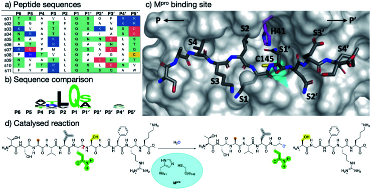
Fig. 1. Substrates processed by SARS-CoV-2 Mpro. (a) The 11 SARS-CoV-2 Mpro cleavage sites, and the corresponding 11-residue peptides, s01–s11; positively/negatively charged residues are blue/red, respectively; histidine is purple; residues with polar sidechains are green; and cysteine is yellow. (b) Comparison between the 11 substrate sequences (generated by WebLogo)9 highlighting the completely conserved Gln at P1 and the highly conserved Leu at P2. (c) View of an energy minimised model, built using apo Mpro (PDB: 6yb7, light grey surface),10 of Mpro complexed with s05 (dark grey sticks); subsites S4–S4′ are labelled. The oxyanion hole formed by the Mpro backbone NHs of Gly-143, Ser-144 and Cys-145 is cyan. (d) The reaction catalysed by Mpro exemplified by s01. Substrate residues important in recognition (see main text) are highlighted.

Fig. 1