. 2021 Sep 6;12(41):13686–13703. doi: 10.1039/d1sc03628a

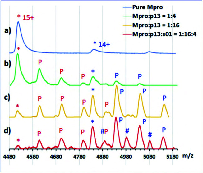
Fig. 11. Non-denaturing MS analysis of designed peptides binding to the Mpro dimer. Inhibitor binding from non-denaturing MS showing normalized intensity in the m/z region around the 14+ and 15+ charge states of the Mpro dimer. (*) indicates unbound Mpro dimer. (a) 5 μM Mpro solution; (b) 4-fold excess of p13 relative to the Mpro dimer; ‘P’ indicates sequential binding of p13 peptides to Mpro in the 15+ charge state (red) and 14+ state (blue); (c) 16-fold excess of p13; (d) 16-fold excess of p13 and 4-fold excess of s01; hash (#) indicates sequential binding of s01-cleavage products (note: the resolution is not sufficient to distinguish between the N- and C-terminal fragments; some non-specific binding of p13 is also observed in (c) and (d) due to the high concentration of the peptide).

Fig. 11