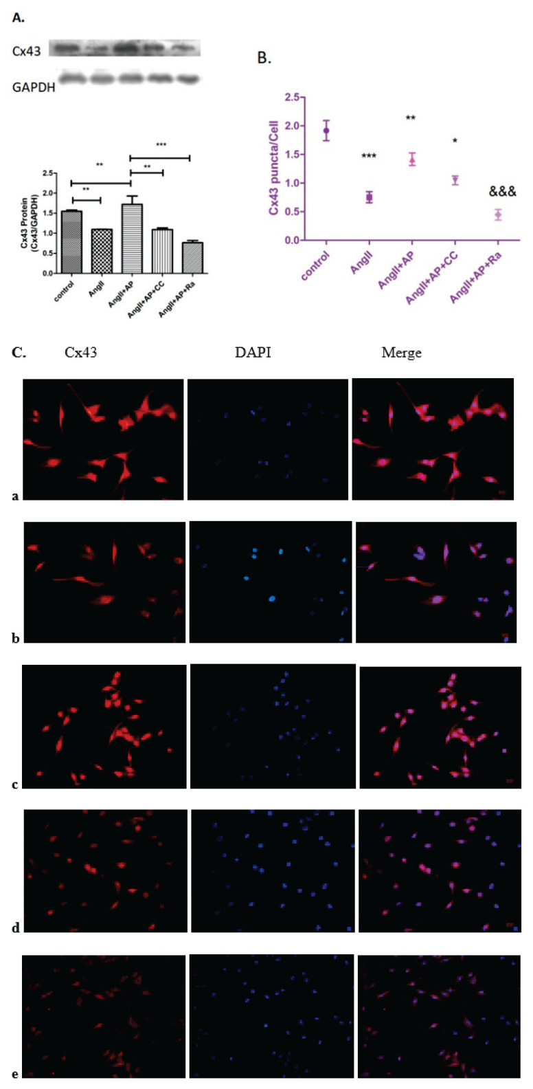
Fig. 3.
The effects of compound C and rapamycin on Cx43 expression and distribution. Cx43 expression and distribution in HL-1 cells was determined by cell immunofluores-cence and Western blot analysis. (A) Compared with the control group, Cx43 expression and distribution in the AngII group was decreased, whereas addition of apelin-13 restored Cx43 expression. Under the treatment of AngII and apelin-13, adding CC or Ra downregulated the expression and distribution of Cx43. (B, C) n=6; * p<0.05 vs. AngII+AP; ** p<0.01 vs. AngII; *** p<0.001 vs. Control; &&& p<0.001 vs. AngII+AP.

