Fig. 1.
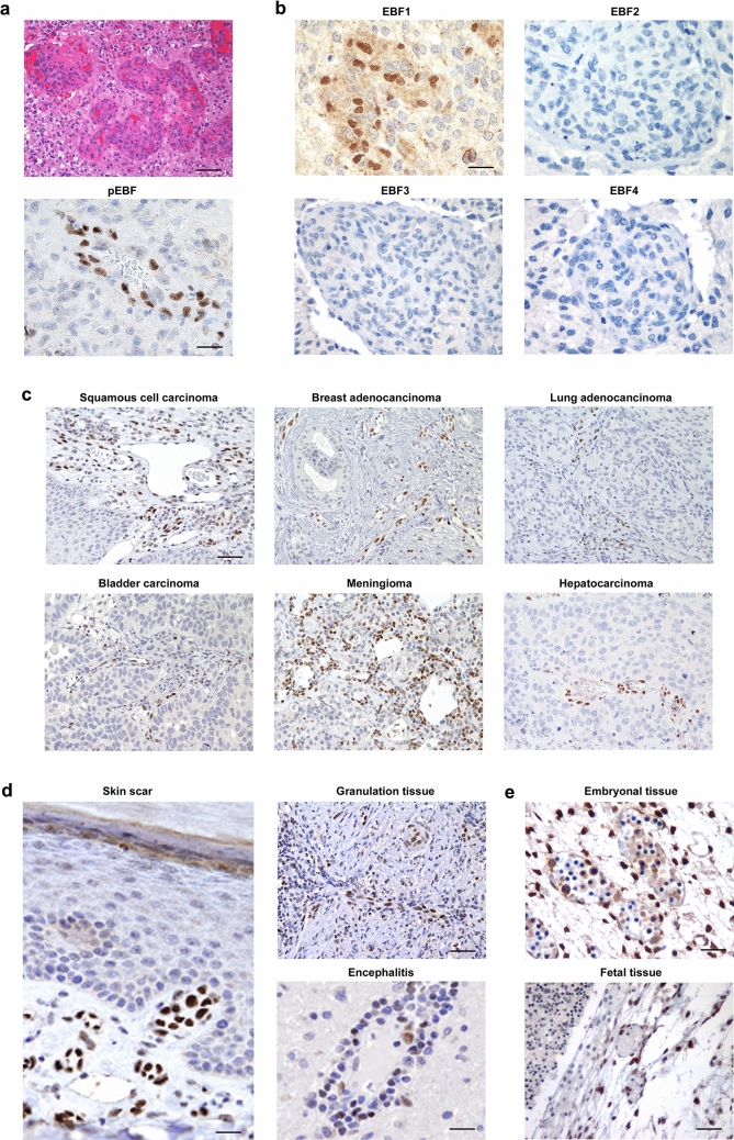
EBF1 is expressed in a cell population within the vessel wall in both neoplastic and non-neoplastic conditions. a Using an anti-pan-EBF antibody, glomeruloid vascular proliferation in glioblastoma (upper image, H&E staining, ×20 original magnification) shows immunoreactivity for EBFs (lower image, anti-pan-EBF immunostain, ×40 original magnification). b When specific antibodies for all the different EBF family members (EBF1–4) were employed, only EBF1 showed positive immunostaining (all images are ×40 original magnification). c Representative sections from different tumor samples show that newly formed tumor vessels selectively express EBF1 independently of their histotype, suggesting that EBF1 expression is a common feature of tumor vessels (all images are ×10 original magnification). d EBF1 is also expressed in the walls of small vessels in physiological and pathological non-neoplastic conditions, i.e. surgical scars (left image; ×40 original magnification), granulation tissue (upper right image; ×20 original magnification) and inflammatory lesions such as encephalitis (lower right image; ×40 original magnification). e EBF1 is strongly expressed in the vessel wall in embryonic (upper image, ×40 original magnification) and fetal tissue (lower image, ×20 original magnification). From panel (c–e) all immunostaining was performed with a specific antibody for EBF1. Scale bars: ×10, ×20 and ×40 original magnification, corresponding respectively to 200 μm, 100 μm and 50 μm

