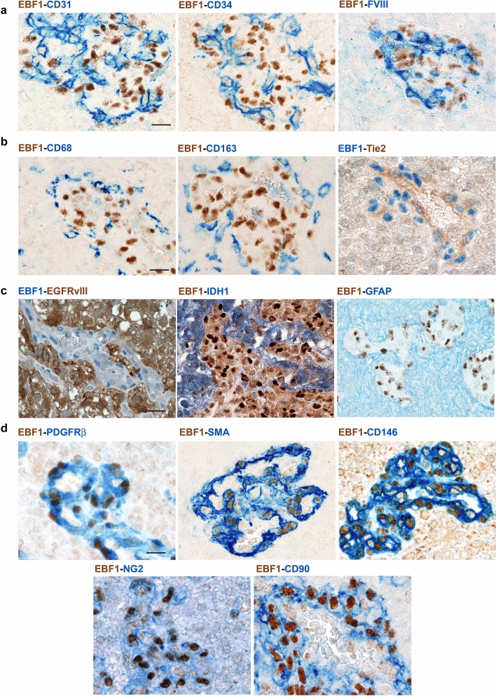
Fig. 2.
EBF1-expressing cells are pericytes. Double immunostaining combining the specific antibody for EBF1 and lineage-specific markers was applied for glioblastoma samples with prominent glomeruloid vascular proliferation in order to identify the phenotype of EBF1-expressing cells. Double immunostaining using endothelial markers CD31, CD34, FVIII showed no double-positive cells (a), and the same was found for monocyte/macrophage markers (CD68, CD163, Tie-2) (b) and for the histological hallmarks of tumor-derived glioma cells (GFAP, EGFRvIII, IDH1-R132H) (c). In contrast, double immunostaining using the most widely recognized mesenchymal/pericyte markers (PDGFRβ, SMA, CD146, NG2 and CD90) (d) revealed the pericyte phenotype of EBF1-expressing cells. Chromogen used to identify immunoreactivity is either brown or blue according to the corresponding label, as indicated. All images are ×40 original magnification; scale bar corresponds to 50 μm

