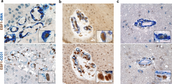
Fig. 3.
EBF1-expressing cells are pericytes. Double immunostaining for EBF1 combined with either pericyte (SMA) or endothelial (CD31) lineage-specific markers on representative tissue sections from other neoplastic lesions (squamous cell carcinoma; left upper and lower images), physiological conditions (skin scar; middle upper and lower images) and inflammatory conditions (encephalitis; right upper and lower images) confirmed the pericyte phenotype of EBF1-expressing cells. Remarkably, the double staining with the endothelial markers clearly highlights the peri-endothelial distribution of the EBF1-expressing cells. In double immunostaining, EBF1 shows nuclear staining, while all the other markers showed cytoplasmic or membrane staining. Chromogen used to identify immunoreactivity is either brown or blue according to the corresponding label, as indicated. Higher magnification of the co-localized areas. All images are ×40 original magnification; scale bar corresponds to 50 μm. Insets are photographic enlargement of a representative field, as indicated

