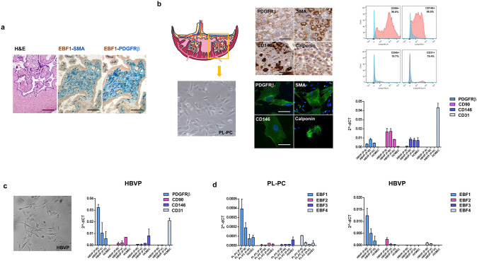
Fig. 4.
EBF1 is expressed in pericytes isolated from both human placenta and brain. a Representative section from human placenta (left image, H&E staining; ×20 original magnification) shows that EBF1-positive cells within the vessel wall have a pericyte phenotype, as previously described. Double immunostaining for EBF1 (brown nuclear) and SMA, PDGFRβ (blue cytoplasmic); ×20 original magnification. b Placental-derived pericytes (PL-PC; left images, ×40 original magnification) were phenotypically characterized by immunocytochemistry on cell blocks (upper middle panels; ×40 original magnification) and immunofluorescence on cultured cells (lower middle panels; ×100 original magnification) using pericyte markers (PDGFRβ, SMA, CD146, calponin), confirming their pericyte phenotype. PL-PCs at passage II (P-II) were also analyzed by flow cytometry using specific antibodies for CD90-APC, CD146-PE, CD45-FITC and CD31-Pe-Cy7 (red). PL-PCs were positive for the pericyte/mesenchymal markers CD90 and CD146 and negative for CD45 (hematopoietic marker) and CD31 (endothelial marker). The negative control is also shown (no antibodies, light blue) (upper right images). RT-qPCR analysis (lower right image) shows that the pericyte phenotype is maintained in culture at least until P-IV, as indicated by the expression of PDGFRβ, CD90 and CD146 and the negativity for CD31. The endothelial counterpart (HUVECs used as control) shows a higher level of CD31 expression, as expected. Of note, CD146 was also detected in HUVECs, in accord with reported data (Du et al. 2015). c Commercially available HBVPs were used as a second pericyte cell line of different embryological origin (neuroectoderm) as compared to PL-PCs (mesoderm) (left image, ×40 original magnification). Their pericyte phenotype was confirmed by RT-qPCR (right image). As expected, HBVPs expressed PDGFRβ, CD90 and CD146, whereas HCMECs, the endothelial counterpart, expressed CD31. HBVPs maintained the pericyte phenotype in culture at least until P-VI even with a progressive decrease in PDGFRβ expression. d The expression of the EBF family members was assayed by RT-qPCR in both PL-PCs and HBVPs and their endothelial counterparts. As expected, both PL-PCs (left histogram) and HBVPs (right histogram) expressed high levels of EBF1, while the expression of the other EBF family members was very low or barely detected. Of note, the absolute expression level of EBF1 in HBVPs was more than 20-fold higher than that of PL-PCs, probably related to their different embryological origin. Scale bars: ×20, ×40 and ×100 original magnifications, corresponding respectively to 100 μm, 50 μm and 20 μm

