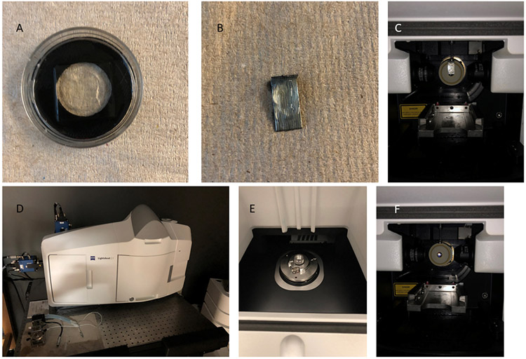
FIGURE 5:
Illustration of sample holders for fluorescence microscopy of mouse brain tissue. (A) Mouse brain slices placed in a glass bottom dish with mounting solution and sealed with a coverslip. (B) A hemibrain mounted on a staple pin and (C) placed inside a light sheet microscope chamber. (D) A light sheet microscope imaging chamber which has (E) a magnet in an upper chamber and (F) a drawer to put the sample and refractive index matching solution in a chamber

