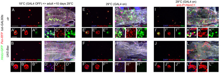
FIGURE 5.
Adult blood cells are still present following the ablation of Hml+ hemocytes. Confocal views of adult bleeds (A,B,E,F,I,J) and abdominal hubs (C,D,G,H,K,L) from tub-GAL80ts, Pxn-RedStinger, HmlΔ-GAL4,UAS-2xEYFP (A,C,E,G,I,K) and tub-GAL80ts, Pxn-RedStinger, HmlΔ-GAL4,UAS-2xEYFP, UAS-Bax (B,D,F,H,J,L) flies expressing (or not) Bax in Hml+ cells only during adulthood (A–D) or throughout their life (E–L). Cells were stained with phalloidin (A,B,E,F: red; C,D,G,H,K,L: white; I,J: no phalloidin) and nuclei with DAPI (blue). (I–L): immunostaining against P1/NimC1 (red). Lower panels show high magnification views of hemocytes. (A’–L’) Merged blue, red and green channels, (A”–L”): red channel only, (A”’–L”’): green channel only. Upper panels: scale bar 200 μm; lower panels: scale bar 20 μm.

