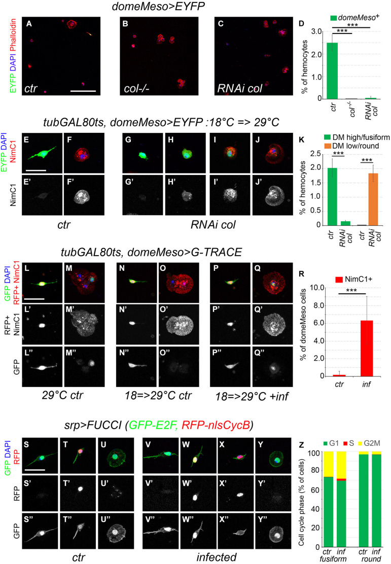
FIGURE 7.
Fusiform cells can differentiate and proliferate. (A–C) Confocal views of blood cells from domeMeso-GAL4, UAS-2xEYFP (A, ctr), col–/–, domeMeso-GAL4, UAS-2xEYFP (B, col–/–) and domeMeso-GAL4, UAS-2xEYFP, UAS-RNAi col (C, RNAi col) adult flies. Blood cells were stained with phalloidin (red) and DAPI (blue). Scale bar 200 μm. (D) Quantifications of the proportion of domeMeso+ hemocytes. Means and standard deviations of at least three independent experiments are presented. A total of 2612, 2350, and 4271 hemocytes were scored in control (ctr), col–/–, and RNAi col flies, respectively. ***p < 0.001 (Student’s t-test). (E–J) Blood cells from tub-GAL80ts, domeMeso-GAL4, UAS2x-EYFP adult flies expressing (G–J) or not (E,F) col RNAi only during adulthood. P1/NimC1 expression (red/white in lower panels) was revealed by immunostaining. Nuclei were stained with DAPI (blue). Scale bar: 20 μm. (K) Quantifications of the proportion of domeMesohigh/fusiform hemocytes (green) and domeMesolow/round hemocytes in tub-GAL80ts, domeMeso-GAL4, UAS2x-EYFP adult flies expressing (RNAi col) or not (ctr) col RNAi during adulthood. Means and standard deviations of at least three independent experiments are presented. A total of 2892 and 4489 hemocytes were scored in ctr and RNAi col flies, respectively. ***p < 0.001 (Student’s t-test). (L–Q) Blood cells from tub-GAL80ts, domeMeso-GAL4, G-TRACE flies raised at permissive temperature (L,M) or switched from restrictive to permissive temperature 24 h after adult emergence (N–Q). domeMeso live expression is revealed by nuclear RFP (red) and its lineage-traced expression by nuclear GFP (green). P1/NimC1 expression (red) was revealed by immunostaining. Nuclei were stained with DAPI. Scale bar: 20 μm. ctr, control non-infected flies; inf, flies infected with E. coli. (R) Quantifications of the proportion of domeMeso+ cells expressing NimC1 in tub-GAL80ts, domeMeso-GAL4, G-TRACE flies raised at permissive temperature only during adulthood and infected (inf) or not (ctr) with E. coli. Means and standard deviations of at least seven independent experiments are presented. A total of 369 (ctr) and 449 (inf) domeMeso+ cells were scored. ***p < 0.001 (Student’s t-test). (S–Y) Representative images of the cell cycle status observed in fusiform or round hemocytes from srpD-GAL4, UAS-FUCCI control (S–U) or E. coli-infected (V–Y) adult flies. Cells in G1 phase express the nuclear GFP only, cells in G2/M express both nuclear GFP and RFP, cells in S phase express the nuclear RFP only. Cell morphology was visualized by phalloidin staining (green). Nuclei were stained with DAPI (blue). Scale bar 20 μm. (Z) Quantifications of the proportion of fusiform or round hemocytes in the different phase of the cell cycle in control (ctr) or in E. coli-infected (inf) srp>FUCCI adult flies. Mean values from at least six independent experiments are presented. A total of 646 (ctr) and 515 (inf) fusiform hemocytes as well as 842 (ctr) and 1336 (inf) round hemocytes were scored.

