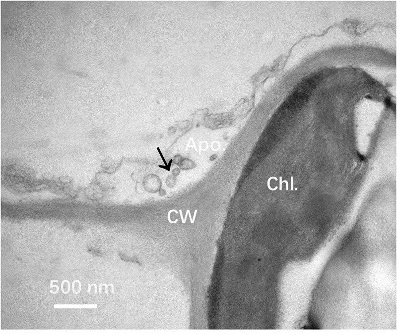
Figure 1.

Electron microscope analysis of Nicotiana benthamiana EVs in leaf tissue. Leaves Nicotiana benthamiana plants were analyzed under a transmission electron microscope. A representative image from three independent repeats is shown. Vesicle-like structures were observed in the apoplastic area (arrow). Apo.: Apoplast; CW: Cell wall; Chl.: Chloroplast. Bar = 500 nm.
