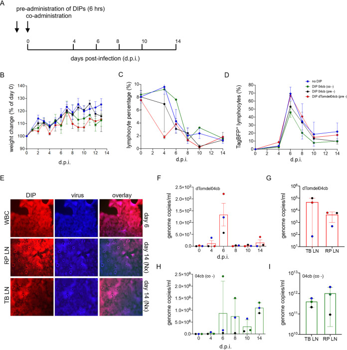
FIG 7.
rCDVRI infection in ferrets. (A) Experimental outline of ferret infection. Ferrets were either pre- or coadministered with DIP nDIRI04cb and preadministered with sDIRIdTomdel04cb or culture medium prior to infection with the challenge virus rCDVRITagBFP. Preadministration was carried out 6 h before infection. (B) Weight change of ferrets during the course of infection. (C) Lymphocyte percentage in EDTA-blood determined by flow cytometry show lymphopenia. (D) Virus infection in lymphocytes was determined for each time point by monitoring fluorescence using flow cytometry. A peak in infection is seen at day 6. (E) sDIRIdTom04cb and virus were isolated from WBC (6 dpi) and from lymph nodes (14 dpi) from ferrets. TB-LN, tracheal-bronchial lymph node; RP-LN, retropharyngeal lymph node. (F and G) sDIRIdTom04cb genome copies were detected in WBC at each time point during the course of the infection and at necropsy in single-cell suspensions of lymphoid tissues. (H and I) nDIRI04cb genome copies were detected in WBC at each time point during the course of the infection and at necropsy in lymphoid tissues.

