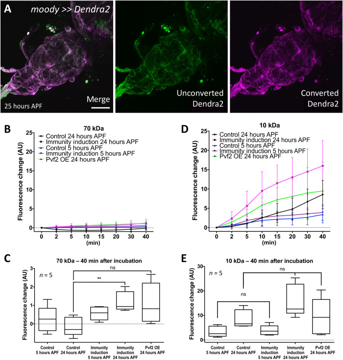
Fig. 7. Immunity induction does not affect BBB integrity.
(A) The BBB-forming cells are not replaced during onset of pupal development. Confocal image of a 24- to 25-hour APF pupal brain. Larvae with the genotype (moody-Gal4 UAS-tub-Dendra2) were subjected to photoconversion of the subperineurial glia covering the ventral nerve cord. The resultant red fluorescent Dendra2 protein can be detected in pupal brain 25 hours APF. Scale bar, 100 μm. (B) Quantification of dye uptake experiments using fluorescein-labeled 70-kDa dextran in control (repo-Gal4, UAS-GFPdsRNA) and after pan-glial immunity induction (repo-Gal4, UAS-PGRP-LE). Datasets were obtained 5 and 24 hours APF. In addition, we included 24-hour APF pupal brains expressing Pvf2. AU, arbitrary units. OE, overexpression. (C) Quantification of changes in fluorescence uptake after 40 min. A slight but significant increase in fluorescein-labeled dextran can be detected following immunity induction (**P = 0.0079) but not following Pvf2 expression (P = 0.0556). (D) Quantification of dye uptake experiments using Texas Red–labeled 10-kDa dextran in control and after pan-glial immunity induction. The same genotypes and time points as in (A) were used. A large variability of data points was found resulting in large error bars. In all genotypes analyzed, an increase in Texas-red–labeled dextran in the CNS can be detected. (E) Quantification of changes in Texas-red uptake after 40 min. A significant increase in Texas-red–labeled dextran was found for control brains (P = 0.0317) and those with a pan-glial immunity induction (P = 0.0079). However, the levels of Texas-red–labeled dextran were not significantly different between control and pan-glial immunity induction (P5hAPF = 0.667 and P24hAPF = 0.095). Likewise, no differences in Texas-red–labeled dextran uptake were noted in 24-hour APF brains expressing Pvf2 (P = 0.944). n = 5.

