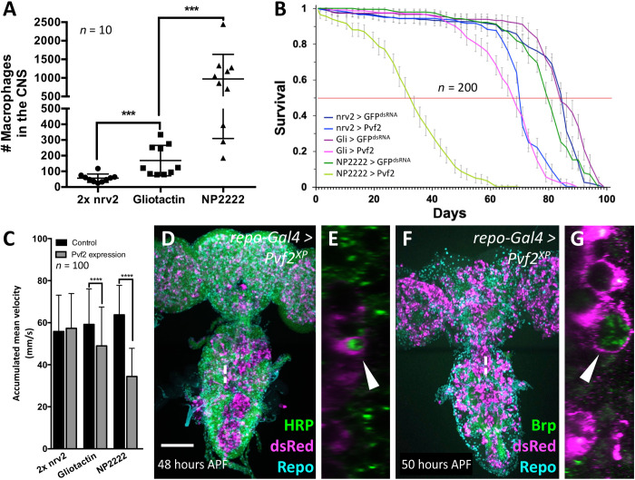
Fig. 9. CNS resident macrophages affect survival and phagocytose neuronal membranes.
(A) Number of macrophages invading the brain of 1-week-old adult flies expressing Pvf2 with different glial cell–specific Gal4 drivers. The P values are ***Pnrv2 vs. Gli = 0.0001 and ***Pgli vs. NP2222 = 0.0002 (Mann-Whitney). (B) Same genotypes as in (A). Longevity of female flies with glial subtype–specific Pvf2 expression compared to control flies. The corresponding Gal4 driver element is indicated in the figure. For control, we expressed GFPdsRNA. The viability is reduced upon Pvf2 expression in all glial subtypes tested and inversely correlates with the number of invading macrophages (n = 200 females in groups of 20 each; ****Pnrv2 = 4.425 × 10⁻³², ****PGli = 5.2984 × 10−41, and ****PNP2222 = 1.9936 × 10−89). (C) Same genotypes as in (A). The climbing ability of 7-day-old females is reduced upon Pvf2 expression and again inversely correlates with the number of invaded macrophages (Pnrv2 = 0.237, ****PGli < 0.0001, and ****PNP2222 < 0.0001). (D to G) Two-day-old pupae with pan-glial Pvf2 expression stained for macrophages using [hml∆dsRed] and subsequent anti-dsRed staining, and either anti-HRP (green) (D and E) to label neuronal membranes or anti-Brp (green) (F and G) to label synapses. Note that macrophages harbor vesicles containing neuronal membrane material (arrowheads). Scale bar, 100 μm.

