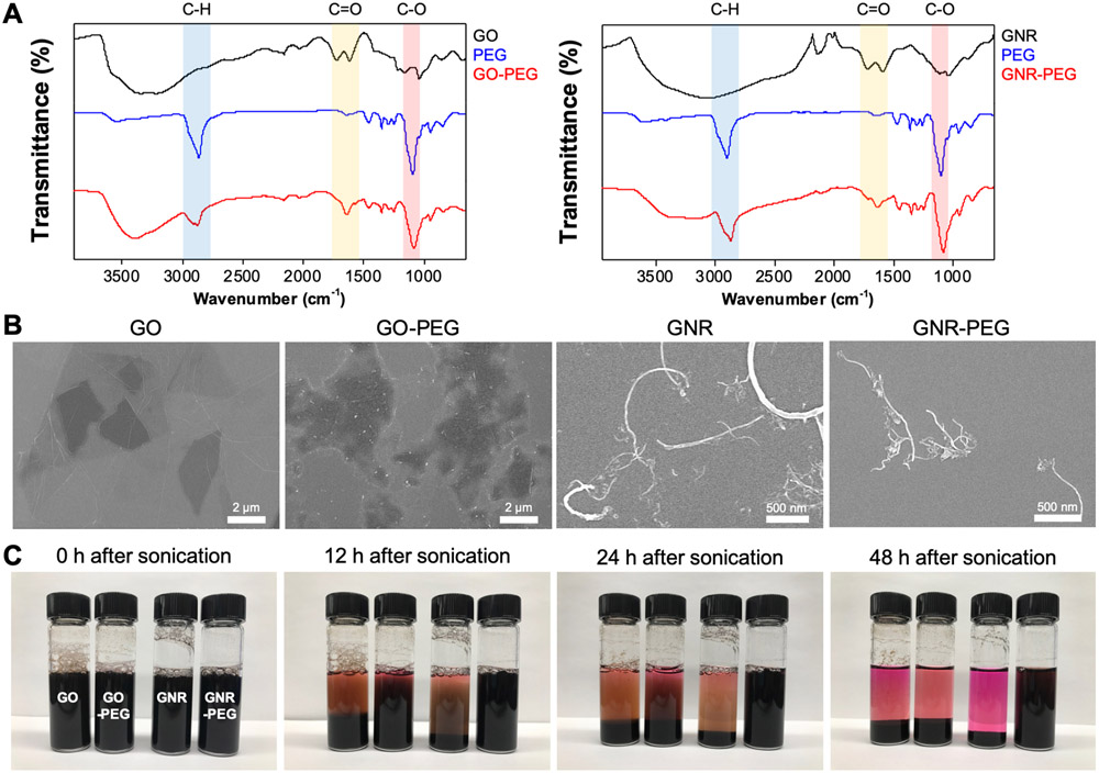
Figure 1.
PEGylation of GO and GNR to reduce aggregation in protein-rich ascites. (A) FTIR spectra of GO-PEG (left) and GNR-PEG (right). Signature peaks of 4-arm PEG were detected in the FTIR spectra of GO-PEG and GNR-PEG: C-O stretch (red region at ~1100 cm−1), COO-H/O-H stretch (blue region at ~2850 cm−1), and C=O stretch (yellow region at ~1640 cm−1). (B) Representative SEM images of GO, GO-PEG, GNR, and GNR-PEG revealed no significant morphological changes after PEGylation. (C) Aggregation test in cell culture media containing 20% (v/v) serum (FBS). Graphene-based materials (100 μg mL−1) settled over time in serum-rich media due to aggregation.

